GSI High-Definition Scientific Digital LED Microscope, USB Video Connection to Computers/Laptops/Notebooks, Magnifies All Microscopic Articles 400x, Special Image Treatment, Includes Stand for Handsfree Use, for Hobby/Science/Education/Industrial- Black Overview
This season discover the Micro world with this newest innovation from GSI. This product was designed by our World-Class Micro experts to capture and display the tiniest miniscule objects in 400 times magnified viewing. Connect to your computer with USB cable included, simply point-and-Snap, and watch the world come alive in front of your eyesGSI High-Definition Scientific Digital LED Microscope, USB Video Connection to Computers/Laptops/Notebooks, Magnifies All Microscopic Articles 400x, Special Image Treatment, Includes Stand for Handsfree Use, for Hobby/Science/Education/Industrial- Black Review
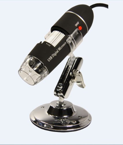

GSI High-Definition Scientific Digital LED Microscope, USB Video Connection to Computers/Laptops/Notebooks, Magnifies All Microscopic Articles 400x, Special Image Treatment, Includes Stand for Handsfree Use, for Hobby/Science/Education/Industrial- Black Feature
- Microscope Lens (25x - 400x), 8 LED Light Source, Snap Function, 5x Digital Zoom
- Color CMOS Image Censor, High Speed CPU, 1280x960 Resolution, 2M Pixels Lens
- USB Compatible for Instant Video Viewing on Computer or Laptop Screen
- Auto White Balance Control, Sharpness Control, Color Mangement, Brightness Adjustable
- High Quality Image Enlarges Microscopic Articles on Skin, Cloth, Leather, Coins, Bills and Hair, Etc.
Related Products
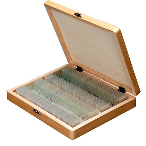
- MICROSCOPE SLIDES 72/pk 1x3", 1mm
- Carson MM-200 Carson Micromax LED 60X-100X LED Lighted Pocket Microscope
- Aven 26700-311 Digital Microscope Universal Stand with X-Y Base and Built in LED Backlight
- Veho VMS-004 Discovery Deluxe USB Microscope with x400 Magnification & Flexi Alloy Stand
- 420 Scope 60-100x LED Handheld Microscope
Customer Reviews
*** Product Information and Prices Stored: Mar 18, 2013 02:30:03
Related : Sun Solar cell hd blu ray player