Friday, November 30, 2012

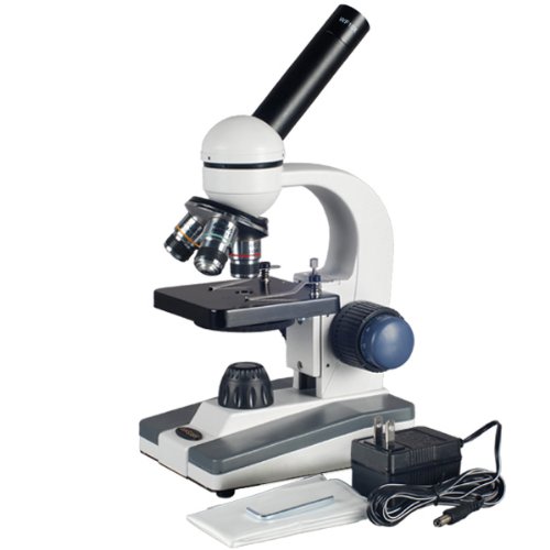
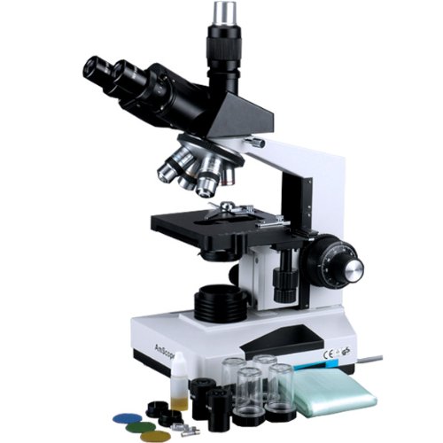
AmScope 20x-60x Student Binocular Stereo Microscope with Top and Bottom Lights


AmScope 20x-60x Student Binocular Stereo Microscope with Top and Bottom Lights Review


AmScope 20x-60x Student Binocular Stereo Microscope with Top and Bottom Lights Feature

  • Widefield Binocular Stereo Microscope Offers Two Stereo Magnification Settings, 20X & 60X
  • High Quality Glass Optical Lenses Provide Sharp Clear Stereo Erect Images
  • All-Metal Framework with Pillar Stand and Both Incident (top) and Transmitted (bottom) Illuminations
  • Great for Explorating the World of Surface Details and Fine Structures; Perfect for Examination of Insects, Plants, Rocks, Stamps, Coins, Jewel, Soldered Points, Microelectronic Components or Virtually any Three Dimensional Objects
  • 5-Year Warranty against Manufacturing Defects Including Parts and Labors

AmScope 20x-60x Student Binocular Stereo Microscope with Top and Bottom Lights Overview

This binocular stereo microscope comes with two magnification power settings (20X & 60X) and a versatile illumination system that provides both incident (top) lighting and transmitted (bottom) lighting. You can choose between incident illumination shining down onto the object or transmitted illumination through the frosted stage plate. The first is used for the observation of three-dimensional objects and the second for the observation of slides. This microscope offers high resolution and good depth within a broad field of view. It gives sharp clear stereo images. Its 45° inclined binocular head ensures an easy observation and rubber eyeguards provide further comfort. This microscope is useful for many applications. It can be used in geology, forestry, archaeology, agriculture and biology. It is appropriate for educational purposes in hospitals and schools. It is great for the exploration of the world of surface details and fine structures of various materials like wood, metal, solder, semiconductor and stone. It is perfect for the examination of insects, plants, rocks, stamps, coins, jewel, soldered points, microelectronic components or virtually any three dimensional objects. This scope is an ideal tool for watchmaker, jeweler, engraver, dies-maker, philatelist, coin collector, students, and etc. It comes with first-class quality and affordable price. It is brand new in factory-sealed box. Its retail value is 0.
*** Product Information and Prices Stored: Nov 30, 2012 23:30:04

Tags : Measurement Guide kodack easy share Men Stainless Steel Watch

Thursday, November 29, 2012

Pathophysiology and Clinical Manifestation of Anemia

The basic physiologic fault caused by anemia is a decrease in the oxygen-carrying capacity of blood and consequently a allowance in the whole of oxygen available to the tissues. Most of the clinical manifestations are directly attributable to tissue hypoxia. Muscle feebleness and easy fatigability are common, although children seem to have a grand quality to function quite well despite low levels of hemoglobin.

The skin is usually pale to a waxy pallor in severe anemia. Cyanosis is typically not evident; because it is the follow of the quantity of deoxygenated hemoglobin and/or red blood cells, not inadequate oxygen saturation of existing hemoglobin. The physician should also keep in mind that skin pigmentation can alter one's estimate of kin pallor.

Light Microscope Images

Central nervous law manifestations inclined headache, dizziness, light- headedness, irritability, slowed notion processes, decreased attention span, apathy, and depression. Increase retardation resulting from decreased cellular metabolism and coexisting anorexia is a common looking in chronic severe anemia. It is oftentimes accompanied by delayed sexual maturation in the older child.

The effects of anemia on the circulatory law can be profound. A allowance is hemoglobin attention that results in decreased oxygen-carrying capacity of the blood is related with a compensatory Increase in heart rate and cardiac output. Initially this greater cardiac yield compensate for the lower oxygen-carrying capacity of the blood, since blood replenished with oxygen returns to the tissues at a faster than general rate.

Diagnostic evaluation
Several tests can be used to the levels of Rbc and hemoglobin. These are habit hematological laboratory procedures. Other tests used to diagnose the basal cause of anemia are included elsewhere in the argument of the single disorder.

Therapeutic management
The objective of curative management is to reverse the anemia by treating the basal cause. For example, in nutritional anemia the exact scantness is replaced. In blood loss caused by hemorrhage, packed red blood cells or whole blood is given. In cases of severe anemia supportive curative care may consist of oxygen therapy recovery of sufficient blood volume, intravenous fluids and bad rest.

Nursing considerations
Since anemia is not a disorder but a symptom of some basal problem, nursing care is related to determining the cause, fostering suitable supportive and therapeutic treatments, and decreasing tissue oxygen requirements.

Assist in establishing a diagnosis. Although, the bodily examination yields necessary evidence regarding the severity of the anemia and some indication of its potential etiology, prognosis primarily rests on hematological blood studies and a faithful history.

Prepare child for laboratory tests. Explicate to older children the need for repeated veni-punctures or finger-sticks for blood analysis. Particularly why a sequence of tests is required. Allow children to play with laboratory equipment and/or partake with test. Older children may enjoy looking at blood smears under a microscope or at pictures of blood cells.

Observe for signs of shock and hypoxia from repeated blood samples. Explicate to parents think for replacing withdrawn blood and necessity of performing tests.

Minimize bodily exertion. Compare child's level of bodily tolerance. Anticipate and help child in those activities of daily living that may be beyond his tolerance. In case,granted diversional play activities that promote rest and quiet but forestall boredom and resignation pick an suitable roommate of similar age and interests and one who requires restricted activity.

Minimize emotional stress. Anticipate child's irritability, short attention span, and fretfulness by contribution to help him in activities rather than waiting for him to ask. Compare parents' awareness of child's need for dependency to conserve strength. Explicate to older children and parents think for behavioral changes caused by anemia. Encourage parents to remain with child.

Place child in room with noninfectious children; restrict visitors with active illnesses. Guidance visitors (and hospital personnel) to practices good hand washing. Narrative any temperature elevation to physician. eye for leukocytosis. Vocalize sufficient nutrition.

Alert ancillary hospital personnel regarding child's bodily tolerance and need for assistance during activity. Keep side rails raised and use safety restraints when applicable.

Be alert to signs of heart failure from excessive cardiac demands of from cardiac over load during blood transfusion. Convention all precautions. Check blood with someone else nurse and physician to ensure spoton blood group/ type with that of child. Run blood moderately and remain with child for infusion of introductory 50ml. Stop blood immediately if any untoward reaction occurs. Attach blood to piggy back setup with general saline or other intravenous solutions to Vocalize open venous line. eye for signs and symptoms of reaction.

Pathophysiology and Clinical Manifestation of Anemia

Related : The Pneumatic Material Handing Products Pneumatics and Plumbing Good choice cheap unlimited cell phone plans Measurement Guide

Wednesday, November 28, 2012

Diamonds for Sale: easy Tips On How To Tell If A diamond Is Real Or Fake

Diamonds for sale on the internet enables men and women around the world to browse among hundreds of styles and designs in the comfort of their homes or offices. One can find numerous websites presenting tons of data as well as astounding images of diamond jewelries plus marvel straight through a lot of consuming finds whenever you're seeing for that excellent diamond ring.

Online marketing and promotion made the diamond stone even more beloved and desired by most citizen who are planning to buy diamonds for sale. There are a lot of reasons why this priceless gemstone is carefully the best gift a man can give to his extra and beloved woman. Due to the advent of online stores, diamond jewelleries have come to be the beloved choice for engagement rings, anniversary and wedding rings. Its unrivaled character and the price it commands somehow equates to the love you have for your partner.

Light Microscope Images

More often than not, men are too lazy to shop around going from one jewelry shop to another in quest for that perfectly designed diamond ring. Searching diamonds for sale online is much easier and a time salvage method. These online jewelry shop are very convenient to use. Being customer oriented a site visitor and prospective clients are provided the full details and data about their chosen diamond rings. Most websites showcase their products straight through images.

Each photograph displays product features and dimensions manufacture it easier to decree on and pick the ring that suits yours and your partner's taste. Moreover, online shopping can also save you money as most of these shops can offer their diamonds for sale at lower prices because they don't pay for space rentals. These online sites can even showcase a lot of varieties much more than an commonplace jewelry shop. The best part is you can spend more time seeing at each photo allowing you more time to collate and pick what you think is the best piece from a lot of options presented.

When you shop for diamond jewelry online the most crucial thing to do is to ask for a certificate issued by a well known diamond grading and skilled appraisers. Almost all appraisers rely on a standardized grading system for diamond stones. Even if you are being issued with a certificate of authenticity from the jeweler it still is not an guarnatee that you have purchased a real diamond.

Read on some helpful tips on how to tell if a diamond is real or fake:

• Check for the refractive properties. A real diamond throws off sparks when light passes straight through it because diamonds possess a high refractive index.
• For loose diamonds, try placing it upside down on a newspaper or any surface, if you can see the bottom then, it's not a real diamond.
• A real diamonds' reflection should be grayish in color not the rainbow reflections that most citizen believe.
• A real diamond shouldn't have an orange flash when checked under a microscope.
• A diamond displays blue fluorescence when settled under Uv light. If you see this color then rest assured that what you got is a real one.

Diamonds for Sale: easy Tips On How To Tell If A diamond Is Real Or Fake

Recommend : Pneumatics and Plumbing Good choice The Fasteners Fasteners Equipment cheap family cell phone plans

Tuesday, November 27, 2012

The Value Of ancient Microscopes

Have you ever met an avid accumulator of aged microscopes? They are as rare as their collections. Some collectors and science museums have microscopes dating back to the 18th century. Zaccharias Janssen invented the microscope nearby 1590. The first real microscope was used in the 17th century by Anthony Leeuwenhoek of Holland. He was intrigued by the way glass lens could magnify things to a great extent and learnt how to make magnifying lenses. With these small pieces of glass he was the first person to be able to see bacteria, yeast and blood cells. He is called the 'Father of microscopy' because of his farranging research and work to improve the capability of these useful scientific instruments in his lifetime.

So now that we know a bit about the history of the microscope, let's see why aged microscopes are considered so valuable. Museums and collectors are willing to pay any amount to get their hands on most of these antiques. The best place for you to go and have a look at them would be in a science museum. Most museums have a huge collection of microscopes to educate people about their history and their significance in modern life.

Microscope

Antique microscopes, as any other antiques, are invaluable because of their uniqueness and rarity. These types are commonly one of a kind as they were not mass produced in earlier centuries. In fact it was only in the middle of the 19th century that associates in Germany and America started yield of microscopes on a large scale. Today, there are many associates in Japan, China and Germany that establish high capability microscopes and sell them all over the world. It's fascinating to note that there are no microscope manufacturing associates in Usa. They import them from other countries.

Most of the aged microscopes were made of brass. They were very sturdy and looked like a telescope mounted on a stand. There were even some binocular microscopes made, which if you ask me look like two telescopes stuck together and mounted on a stand! You cannot fantasize how many dissimilar variations of the microscope were made in the early years before the modern type was created. These big brass microscopes came with many accessories like lenses of dissimilar variations, slide preparation tool and other parts. If you own one of these microscopes along with its accessories, it would be considered priceless today.

These types of old and primary microscopes are collected by many who feel the need to keep these first-rate pieces. They are collected so that our children and their children can see how these instruments were made and used hundreds of years ago. These are things we take for granted these days but it took many years of research and experimentation before great scientists were able to establish and improve upon the microscope. Today a microscope is used for so many things. It is leading for scientists to show the way their research in developing new medicines for us. Doctors take their help to diagnose diseases.

There are so many dissimilar types of microscopes made and used today. They are used in research laboratories, in medical laboratories and in schools. The precedents of these modern microscopes help us to learn how they were made and how they have become such an leading part of our lives. aged microscopes will always be a priceless and invaluable part of our history.

The Value Of ancient Microscopes

Recommend : Picking Safety Products Measurement Guide Rackmount Shelves Cell Phone Backup

Monday, November 26, 2012

AmScope 40X-1000X All-Metal Student Compound Microscope with Mechanical Stage and Coarse & Fine Focus

AmScope 40X-1000X All-Metal Student Compound Microscope with Mechanical Stage and Coarse & Fine Focus Review


Prices as of : Nov 26, 2012 09:15:04
Listprice : $500.00
Offier Price : $89.98
You Save : $410.02

AmScope 40X-1000X All-Metal Student Compound Microscope with Mechanical Stage and Coarse & Fine Focus Feature

  • Cordless Monocular Offering Five Widefield Magnification Settings up to 1000X
  • Optical Glass Lenses; Single Lens Condenser; Disc Diaphragm
  • All-Metal Framework; Mechanical Stage with Coaxial Coarse & Fine Focusing
  • LED Illumination Using either Wall-power or Three AA Batteries
  • Wall-power Adapter/Recharger Included

AmScope 40X-1000X All-Metal Student Compound Microscope with Mechanical Stage and Coarse & Fine Focus Overview

This is a perfect microscope for students to learn science, offering five magnification settings, 40X, 100X, 250X, 400X and 1000X. It comes with full optical glass elements, metal framework, 360° rotatable monocular head, coarse & fine focusing, mechanical stage and the LED illumination system that uses either wall-power (adapter included) or three AA batteries (or re-chargeable batteries). It is an ideal microscope for home school or for students from elementary to high school. It is also an excellent instrument for hobbyists. It is brand new in original box. Its retail value is 0.

*** Product Information and Prices Stored: Nov 26, 2012 09:15:04

Recommend : Best Fasteners Sun Solar cell Picking Safety Products led microscope light Moblie Mount

Sunday, November 25, 2012

Veho VMS-004 Discovery Deluxe USB Microscope with x400 Magnification & Flexi Alloy Stand

Veho VMS-004 Discovery Deluxe USB Microscope with x400 Magnification & Flexi Alloy Stand Review


Prices as of : Nov 25, 2012 04:15:05
Listprice : $99.99
Offier Price : $59.56
You Save : $40.43

Veho VMS-004 Discovery Deluxe USB Microscope with x400 Magnification & Flexi Alloy Stand Feature

  • 2.0 Megapixel OMOS lens
  • Up to x400 digital USB microscope with snapshot and video capture and measurement software
  • On/Off LEDs for object illumination
  • Alloy Stand and Stand alone Capture Button
  • Tacton Rubber Touch Housing
  • Pc and Mac Compatible

Veho VMS-004 Discovery Deluxe USB Microscope with x400 Magnification & Flexi Alloy Stand Overview

Handy desktop 400x magnifying USB Microscope

*** Product Information and Prices Stored: Nov 25, 2012 04:15:05

See Also : Picking Safety Products philips hd blu ray player

Saturday, November 24, 2012

Great Price AmScope SM-4TZ-144-5MT for $699.98

AmScope 3.5X-90X Zoom Magnification Stereo Microscope on Boom Stand + 144 LED Light + 5MP USB Digital Camera Review


AmScope 3.5X-90X Zoom Magnification Stereo Microscope on Boom Stand + 144 LED Light + 5MP USB Digital Camera Feature

  • 3.5X-90X zoom magnification power
  • Super widefield of view 2-1/2" (65mm) and large working distance 8" (200mm)
  • 5MP camera captures still images and steams live video on your PC screen
  • Advanced editing, processing and measuring software compatible with Windows 2000/XP/Vista/7
  • Powerful 144 LED bulb ring light

AmScope 3.5X-90X Zoom Magnification Stereo Microscope on Boom Stand + 144 LED Light + 5MP USB Digital Camera Overview

This is a trinocular 3.5X-90X zoom stereo boom microscope with 5M (2592x1944) pixel high resolution USB2.0 color digital camera. It comes with a trinocular head, 30mm SUPER widefield high-eye point eyepieces, a 3D double-arm heavy-duty boom stand, and a 144-LED ring light with dimmer. This microscope offers crystal clear images, super widefield viewing, very large magnifying zoom range (26:1) and a large working distance (8", 200mm). Its high-eye point optic design ensures an easy and comfortable observation, including eyeglass wearers. The color digital camera captures 10.7 MP high resolution microscope images and displays them as full-screen live video on your computer screen. The reduction lens included displays the same field of view images on your computer screen as those seen through the microscope eyepieces. With the included user-friendly software, which is compatible with Windows 2000/XP/Vista/7, you can edit microscope images in a manner similar to PhotoShop. You can conduct measurements across images, record videos, share images, and save them in BMP, TIFF, JPG, PICT, PTL and other formats. You can conduct measurements across microscope images, measuring lengths, angles, areas and etc.The 3D double-arm boom stand allows you to turn the microscope head around three different axis (x, y, and z), allowing you to point the microscope head to ANY direction you wish. The intensity-adjustable LED ring light provides bright, even and cool light illumination.This microscopy system is an ideal instrument for gemologists, engravers, collectors and anyone in semiconductor and/or electronic industry. It is also a right microscope for biological laboratory applications that require low heat release.Made by the same OEM supplier that provides optical instruments for Leica, Zeiss, Nikon and Olympus, it arrives brand new in a factory sealed box and retails for more than ,000.

Related Products

Customer Reviews


*** Product Information and Prices Stored: Nov 24, 2012 02:00:03

My Links : The Fasteners microscope light

Thursday, November 22, 2012

OMAX 10X-20X-40X-80X Digital Binocular Stereo Microscope with Dual Lights System and 2.0MP USB Digital Camera

OMAX 10X-20X-40X-80X Digital Binocular Stereo Microscope with Dual Lights System and 2.0MP USB Digital Camera Review


$356.05

OMAX 10X-20X-40X-80X Digital Binocular Stereo Microscope with Dual Lights System and 2.0MP USB Digital Camera Feature

  • Model: XG223AWF20C20C
  • Head: 45degrees;Inclined binocular with locked-in eyepieces; Eyepieces: wide field WF10X/20 and WF20X; Rotational objectives: 2x, 4x; Magnification levels: 20x, 40x, 80x; Working distance: 2-1/4inch (57mm); Field of view: 2.5mm, 5mm, 10mm; Left ocular-tube diopter adjustment +/-5dp; Interpupillary distance: 2-3/16inch ~ 2-15/16inch(55-75mm)
  • Incident (upper) illuminator: 12V/10W tungsten; Transmitted (lower) illuminator: 12V/10W tungsten; Easy illumination mode switch: [upper] [off] [lower]; Adjustable focus knob tension; Frosted glass stage plate: 3-3/4inch (95mm) in diameter; White/black plastic stage plate: 3-3/4inch (95mm) in diameter; Head vertical moving range (along the pillar): 2-3/4inch (70mm); Focusing adjustment range: 1-5/8inch (42mm); Power supply: 110V/60Hz (US and Canada)
  • Resolution: true color 1600 x 1200 pixels; Interface: USB 2.0 (480Mb/sec); Pixel size: 2.8 um x 2.8 um; Image sensor: 1/3.2inch CMOS; SNR: 42.3 dB; Mounting port: insert it into eyepiece tube of microscope directly; Platform compatibility of camera: Windows 2000(SP.), Windows XP, Windows VistaWindows 7 32/64-bit; USB cord length: 6 feet (183cm); Software: live viewing, video recording, image capturing, measurement, and etc.
  • Dimension: 9inch x 4-3/4inch x 14-3/4inch (23cm x 12cm x 40cm); Net weight: 6 lb 9.5oz (2.990kg)

OMAX 10X-20X-40X-80X Digital Binocular Stereo Microscope with Dual Lights System and 2.0MP USB Digital Camera Overview

Packing List: Binocular stereo microscope with a pair of WF10X and WF20X eyepieces;Frosted glass objective stage plate;Black/white plastic stage plate;Rubber eyeshields;Wrench for focus knob tension adjustment;Allen key for stage lock;Dust cover;Extra tungsten bulb (12V/10W 6F);2.0MP USB camera;0.45X medium lens;CD (camera drive and software);USB cable;30.5mm eyetube conversion adapter

*** Product Information and Prices Stored: Nov 22, 2012 21:00:03
$236.95

Visit : Sun Solar cell Finishing Products wd media player Rack frames 19 inch

Wednesday, November 21, 2012

What is Upper Endoscopy and Why Is Small Intestine Biopsy Recommended for Celiac Disease?

When undergoing an assessment for potential celiac disease or gluten sensitive enteropathy doctors commonly recommend an upper endoscopy and small intestine biopsy. What that may mean or why it is recommended may not be clear to population who are facing the decision to undergo the policy themselves or to branch their child to the exam.

Endoscopy in celiac: What is it and how is it done?

Microscope

The medical name for upper endoscopy is esophagogastroduodenoscopy or Egd for short. The endoscope is a thin flexible tube about the diameter of a fat pencil that has a video chip in the end and channels for flushing of water, suctioning of secretions and duct of instruments. It has dials that allow the tube to be turned up/down and right and left at the tip permitting it to be passed straight through the mouth, down the esophagus or feeding tube, into the stomach and then into the first part of the small intestine the duodenum, hence the name Egd.

Endoscopy in celiac: Do you feel it or remember it?

People undergoing the exam in the U.S. Typically are sedated with a medication. Medications similar to valium with good amnesia and relaxing effect called midazolam or versed combined with a narcotic like meperidine (demerol) or fentanyl are generally used. More recently a very short acting intravenous sedative, propofol (diprovan), may be administered for deep sedation or an intravenous form of normal anesthesia. Occasionally, commonly in very young children or population with severe lung problems, normal anesthesia is required. The exam is commonly not felt or remembered because of the medications.

Endoscopy in celiac: What is examined in celiac and how well can the lining be seen?

Celiac disease affects the upper quantum of the small intestine, in the two sections known as the duodenum and jejunum. The exam of the small intestine is commonly wee to the first section termed the duodenum though occasionally the second section known as the jejunum may be reached especially when a longer endoscope is used. The resolution of video images are very high with the newest endoscopes and also may have a magnification and color inequity mode to detect very subtle signs of damage of the small intestine.

Endoscopy in celiac: What are the typical findings?

The characteristic appearance of the face of the small intestine in celiac disease contain superficial ulcerations that are generally linear, flattening of the folds, notching or scalloping of the folds and a mosaic like pattern. However, the face may appear normal and only under wee exam of samples will the lining show signs of gluten caused injury.

Endoscopy in celiac: What are biopsies?

Samples of small intestine are obtained with biopsy pliers that consist of tiny jaws with cups that permit pinching off samples of the intestinal lining. This is painless and very safe. The samples are sent to a prognosis lab in a preservative solution, processed, mounted on a microscope slide, and stained for exam under the microscope by a pathologist. Small intestine injury from gluten may be patchy, therefore, any samples are recommended. A minimum of 4 pieces and preferably 8-12 samples should be obtained to avoid missing wee signs of celiac disease.

Endoscopy in celiac: What does the pathologist look for on the slides?

The pathologist examines the slide for evidence of damage or injury characteristic of gluten sensitivity. Occasionally special stains are required to see signs of irritation known as inflammation characterized by an increased estimate of a type of immune active white blood cells called lymphocytes. In early celiac and gluten sensitivity without celiac disease the biopsy may be normal and the prognosis cannot be established by the biopsy.

Endoscopy in celiac: Summary.

The policy of endoscopy is safe, painless, and very helpful for establishing the prognosis of celiac disease while excluding other upper intestinal disorders. The main drawback of endoscopy is that nearly every person must have sedation to tolerate the exam and it can be expensive if not fully covered by insurance. Sometimes, celiac disease is diagnosed by endoscopic biopsy in population who either have normal blood tests or as an incidental finding in those undergoing endoscopy for other reasons. Fear or blurring about endoscopy should not forestall anything who is suspected of having celiac or gluten sensitivity from undergoing endoscopy. Supplementary data about celiac disease and other digestive diseases are ready at http://www.thefooddoc.com, the premier website under amelioration by "the food doc", Dr. Scot Lewey, a practicing stomach and intestinal devotee (gastroenterologist).

Copyright 2006, The Food Doc, Llc All rights Reserved. Http://www.thefooddoc.com

What is Upper Endoscopy and Why Is Small Intestine Biopsy Recommended for Celiac Disease?

Tags : The Fasteners Measurement Guide Measurement Guide

Tuesday, November 20, 2012

Microscope Parts - What Is Inside the Microscope?

The most basic type of optic microscope is the magnifying glass. It is best to know all the different microscope parts before purchasing a microscope so that you can select the one that is best for you.

Eyepiece Lens - This is the part of the microscope that the user for real uses to look through onto the object that is being magnified. The eyepiece lens is typically the same in most microscopes except for the children's microscope when the eyepiece lens will be built in a smaller form in order to accommodate a smaller viewers face proportions.

Microscope

Objective Lenses - The objective lenses are the second set of lenses in a composition microscope. There are usually a collection of strengths of objective lenses on any one given microscope. The objective lens will usually range in magnification power from 4X to 100X. When the objective lens is combined with the power of the eyepiece lens, then the magnification power is times by the amount of the two lenses combined. So if your eyepiece lens is 10X in power and you integrate that with your objective lens which is 100X in strength, then you will get the magnification power of 1000X.

Tube - The tube is responsible for connecting together the eyepiece lens and the objective lens. This is a very basic part of the microscope and only serves one singular purpose. The functionality of this piece will usually never vary other than connecting the two lenses and occasionally it will also house some type of adjustment knob on the microscope.

Arm - The arm is responsible for supporting the tube and allowing the tube to be related to the base of the microscope. The arm has no other function other than supporting the tube.

Stage - This is the place where you for real place your slide onto. The stage usually has two clips on it that are used to procure the slide to the exterior of the slide. Some microscopes have a mechanical stage which allows the user to for real move the slide around using knobs instead of manually positioning the slide by hand.

Revolving Nosepiece (Turret) - This piece is used to rotate many objective lenses on one singular microscope. This is used when you need to elect the amount of magnifying power that you want to use.

Rack Stop - This part of the microscope is used to preclude the user from bringing the objective lens down to close to the slide that it breaks or damages something. This setting is set at the premise and cannot be adjusted.

Those are the most basic and common parts of a pocket microscope and you should take the time to check out each one before purchasing your microscope. Knowing all the parts of the microscope is a good way to shape out which one will work best for you.

Microscope Parts - What Is Inside the Microscope?

Thanks To : Sun Solar cell Picking Safety Products The Fasteners easy share digital camera

Monday, November 19, 2012

Digital Microscopes in the Elementary Science Classroom - Effective, Affordable, and Easy to Use

Advances in digital microscope technology over the last few years have resulted in great benefits for students. While there are many distinct types of digital microscopes, they fall into one of two main categories, those that join together to the Tv and those that join together to the computer through a Usb port. Instead of students being required to share microscopes and discuss their findings without any references, the digital microscopes allow the whole class to view specimens and discuss findings as a group.

One type of digital microscope plugs into a data projector or television. This makes the process of teaching science to a classroom much more dynamic, easier and more cost effective. The presentation of specimens and consulation can be done as a matter of fact by placing the television in the front of the classroom where all students have a clear view of the screen. The instructor places the specimen under the handheld microscope for view by the whole classroom.

Microscope

The instructor is able to use the television to point out details of the specimen, encourage participate by all students, and fulfill the objectives of the lesson plan using one microscope. There is no need for software or extra tool to make the microscope a vital part of the educational process.

Students will be able to learn the steps for dissecting specimens properly without the normal trial and error that often accompanies this process. In addition, group discussion, questions, and answers all the time serve educators well when presenting new or unusual content to students. Teaching students about the cellular buildings of animals and plants can be as a matter of fact done using the large, clear, and crisp photo in case,granted by the television screen or data projector.

An added advantage of this type of law is that students with special needs, who may not normally be able to handle the small controls of a microscope or be able to navigate the intricacies of the scope, will be able to participate and learn as a matter of fact with the rest of the class. This inclusion of all students in the science exploration process will improve and empower students with special needs to participate in studying more actively.

The scopes made for use with televisions or data projectors lack the functions and features of the scopes that concentrate with a computer using a Usb port. These systems are able to report still images, do time-lapse videos, and contribute a continual flow of facts to students relative to the specimen they are studying. An instructor can as a matter of fact design a lesson plan that will comprise the splitting of a cell, or the increase process of fungi or bacteria and treat students to the breathtaking excitement of watching nature in action.

The most productive microscopes for study use are designed to contribute greater magnification of 10x to 200x or higher. They also comprise easy-to-use controls for Led lighting which will allow for adaptation to light sources in the classroom more easily.

Many teachers find that using the digital microscope in conjunction with desk microscopes allows for more flexibility in teaching techniques and methods. It is very distinguished to have the instructor be able to display what the students are seeing for in their desktop microscopes. The interaction of students and instructor when students know what they are seeing at allows the instructor to focus on the task and specimen at hand for the whole class instead of spending time at individual desks telling students the same information.

The benefits of using a digital microscope in elementary science classrooms are tremendous. The SmartScope by SmartSchool Systems is affordable and easy to use and opens doors for educators and students. Educators are able to generate dynamic lesson plans that utilize the full applications of the microscope while the students advantage from the breathtaking world that has been opened to them through this modern technology.

Digital Microscopes in the Elementary Science Classroom - Effective, Affordable, and Easy to Use

Thanks To : Best Fasteners Men Stainless Steel Watch olympus light microscope

Saturday, November 17, 2012

Celiac Disease Biopsy Explained: Part I Villous Atrophy

The determination of celiac disease is confirmed by a characteristic abnormal appearance of the small intestine under the microscope. Flattening of the general finger like projections called villi accompanied by signs of inflammation is taken to indicate damage or injury from the storage protein gluten in wheat and similar proteins in barley and rye. The small intestine biopsy has became the gold thorough for establishing the determination of Celiac disease or gluten sensitive enteropathy. Before 1960 gluten resignation followed by revision and subsequent worsening upon rechallenge was the diagnostic criteria.

Early in the 1960's straight through the 1970's the small intestine was biopsied by having population swallow a small metal capsule that was attached to a suction tube. This was used to suction up tissue into the capsule before guillotining off some tissue once the capsule was confirmed to be in the small intestine by x-ray. Now the tissue is obtained by upper endoscopy, the tube of a lighted video scope straight through the mouth under sedation to the small intestine, where biopsies are obtained with cupped forceps.

Microscope

Celiac disease biopsy: What does the pathologist look for under the microscope?

The small intestine ordinarily has finger like projections called villi that give it a large covering area or sense area for absorption. The villi follow in a shag rug or terry cloth towel type appearance. Lining the covering covering of each villous are intestinal cells or enterocytes that secrete mucus and Ant. Eject fluids, nutrients, minerals like iron, and vitamins like B12. On the covering of the enterocytes are digestive enzymes like lactase that Ant. Eject lactose or milk sugar. At the base of the villi are crypts or circular like collections of intestinal cells.

Celiac disease biopsy: What is villous atrophy?

Normally, villi are 3-5 times longer than the crypts are tall. However, intestinal injury can follow in blunting, shortening (partial villous atrophy) or perfect loss of the villi and flattening (villous atrophy) of the intestinal surface. The shag rug will have bare spots or the terry cloth towel becomes like a tee shirt. The follow is lack of absorption of nutrients and water resulting in weight loss, malnutrition, and diarrhea.

Celiac disease biopsy: What if the biopsy does not show atrophy or partial atrophy?

If the villi are at least 3 times as long as the crypts are tall then no flattening or blunting of the villi is gift and celiac disease becomes more difficult for the pathologist to diagnose without the history or blood test results. However, an increased whole of Iel's (intra-epitheliel lymphocytes) in the setting of a obvious exact blood test for celiac, symptoms and especially if supported by proximity of Dq2 and/or Dq8 gene pattern, is highly suggestive of celiac disease. The strangeness comes when the blood tests for the exact tests are negative or not elevated but only the "non-specific" blood tests (anti-gliadin or Aga and anti-reticulin antibodies) are elevated. Also, some population with milder forms of celiac have no blood tests abnormal but have first-rate biopsy findings of celiac and are termed seronegative (blood test negative) celiacs.

Celiac disease biopsy: Can the biopsy be general in celiac disease?

By definition, the biopsy has been carefully the gold thorough for diagnosing celiac. However, recent studies have shown that the biopsy can be general in some population with celiac. How can this be? The pathologist reading the biopsy may illustrate the biopsy as general based on his or her bias about celiac disease, a failure to appreciate the point of the proximity of Iel's, or misuse of the older thorough of >40 Iel's per 100. However, more importantly is the recent recognition that general appearing biopsies may not be normal. Electron microscopy has revealed ultra-structural abnormalities in apparent general biopsies of population confirmed to have celiac disease. Extra stains, that consist of immune labeling of lymphocytes, have also confirmed increased numbers of obvious types of exact lymphocytes in the villi of intestinal biopsies of population confirmed to have celiac. The lowest line is that a general biopsy does not definitively exclude celiac disease or gluten sensitivity.

Celiac diasease biopsy: What are other possible causes of biopsy changes that mimic celiac disease?

Cow's milk protein sensitive enteropathy (Cmse), viral or bacterial infections, medications (especially aspirin like arthritis medications e.g. Ibuprofen etc), autoimmune enteropathy, Helicobacter pylori infection (the stomach ulcer bacteria), Aids, base variable immunodeficiency, and lymphoma of the intestine are all possible causes of small intestine changes that may mimic celiac. However, if you have first-rate celiac type symptoms, a obvious celiac exact antibody (anti-endomysial antibody or tissue transglutaminase antibody) and a obvious response to a gluten free diet then celiac is the likely cause. The likelihood is supplementary increased if you carry one or both of the two major genes linked with celiac disease, Dq2 and/or Dq8. Normalization of celiac exact blood tests and the biopsy after a gluten free diet confirms the determination of celiac disease.

Celiac Disease Biopsy Explained: Part I Villous Atrophy

See Also : Fasteners Equipment Pneumatic Plumbing Cell Phone Backup full hd blu ray players

Friday, November 16, 2012

AGPtek® Black USB Digital Microscope 2 Mega Pixel Video Camera Microscope 20-200X

AGPtek® Black USB Digital Microscope 2 Mega Pixel Video Camera Microscope 20-200X Review


Prices as of : Nov 16, 2012 02:00:05
Listprice :
Offier Price :
You Save :

AGPtek® Black USB Digital Microscope 2 Mega Pixel Video Camera Microscope 20-200X Feature

  • Use in Inspection dissection/examination, Plant dissection/examination, Skin examination, Scal examination, Textile Inspection, Jewelry Inspection, Collections/Coin Inspection, Printing Inspection, PCB or PCBA Inspection and so on.
  • Image sensor: 2 Mega Pixels. Video Capture Resolution: 160x120,320x240,640x480,1280x1024. Still Image Capture Resolution: 160x120, 320x240,640x480,1280x1024.
  • Exposure: Auto. Photo format: JPEG. Light source: 8 LED. PC interface: Mini USB1.1&2.0.
  • Power source: 5V DC from USB port. Gray scale: Level 8. Sleep current:£¼1 mA. Work current:£¼180 mA. Magnification Ratio: 20x to 200x.
  • Operation system: Windows XP, Vista, Win7, Mac OS X 10.5 or above. Color£ÂºBalck.

AGPtek® Black USB Digital Microscope 2 Mega Pixel Video Camera Microscope 20-200X Overview

Description:
USB Digital Microscope is a good quality to use in Inspection dissection/examination, Plant dissection/examination, Skin examination, Scal examination, Textile Inspection, Jewelry Inspection, Collections/Coin Inspection, Printing Inspection, PCB or PCBA Inspection and so on.
The microscope is made up of high-quality IC and electronic components,with clear image quality, low power consumption, high-resolution, applies to small objects to be larger£¬photo and video production, is the best choice of small objects recognization.

Product Features:
1. Image sensor: 2 Mega Pixels.
2. Video Capture Resolution: 160x120, 320x240,640x480,1280x1024.
3. Still Image Capture Resolution: 160x120, 320x240,640x480,1280x1024.
4. Exposure: Auto.
5. Photo format: JPEG.
6. Light source: 8 LED.
7. PC interface: Mini USB1.1&2.0.
8. Power source: 5V DC from USB port.
9. Gray scale: Level 8.
10. Sleep current:£¼1 mA.
11. Work current:£¼180 mA.
12. Save temperature: -20¡Ã£C to +60¡Ã£C.
13. Work temperature: 10¡Ã£C to +40¡Ã£C.
14. Magnification Ratio: 20x to 200x.
15. Operation system: Windows XP/Vista/ Win7/Mac OS X 10.5 or above.
16. Color: Balck.

Package Included:
1 x USB Microscope
1 x Stand
1 x CD Driver


*** Product Information and Prices Stored: Nov 16, 2012 02:00:05

Friends Link : Pneumatics and Plumbing Good choice Pneumatic Plumbing Rackmount 19 inch

Wednesday, November 14, 2012

overview of the modern aggregate Light Microscope

The mixture light microscope is one of the most generally used types of microscope. Among its many uses is the science classroom, where they are an affordable and easy to use instrument for learning biology and chemistry. The instrument traces back generations, but recently has evolved into a very contemporary instrument.

Only a few decades ago, you could find the microscope in just two basic types. There was the monocular mixture microscope and the binocular compound. That is, the mixture light microscope came with whether one or two viewing units. With the monocular compound, there was a singular eyepiece and so you looked at your specimen with just one eye. This is still what many people envision when they hear the word "microscope." Then there's the binocular model which, naturally, resembles a pair of binoculars. That's because there are a pair of viewing units, one for each eye. Today, there are few if any monocular mixture microscopes made. The new standard is the binocular.

Microscope

However, even more modern times have introduced to us a newer standard. This is the trinocular mixture microscope. The "tri" signifies a third viewing port. This makes it a not-as-common spin-off of the binocular compound.

The function of the trinocular microscope is a bit different, since most people only have two eyes. The third eyepiece, then, is for use by a second person. It comes in handy in a combine of situations. First, if two people need to consult together over the same specimen, they are able to look at it at the same time. A second and more common use of the trinocular is to allow a science educator to see the same thing that his learner sees under the microscope. A third good use for it is to take pictures of the specimen with a camera placed over the third eyepiece. Each of the microscope parts functions in harmony, allowing the camera to see what the person's eye sees.

The mixture light microscope, as recommend earlier, is marvelous due to its pair of optical parts. Let's discuss these and how each of them is used.

1. First of these parts are the objective lenses on the scope. These lenses sit right above the specimen, collecting the light emitted by the specimen. The typical mixture microscope has 3, 4 or 5 objective lenses. Each of these will offer a slightly separate level of magnification. These objective lenses are held together by the nose piece. As this nose piece is notated, the magnification is altered by a separate objective lens.

2) The eyepiece is the second major part. This is the part that your eye looks directly straight through to see the specimen. It's a cylinder shape and contains a lens. The eyepiece is also called the microscope ocular.

As these two major parts work together, there are is a huge range in magnification available. In fact, if it fits on the microscope, there is approximately de facto a magnification level that will bring it to life for you. It's virtually unlimited in the viewing possible it offers you.

The mixture scope has some other leading parts, too, like the condenser. The condenser is one of the more fragile but leading parts. There are other leading parts to the microscope, such as the condenser. A diagram of the parts condenser is helpful to make sure it stays in good condition. Do a Google or Yahoo quest for more information.

Costs for your microscope might vary, but regularly they will run in the range of 0 to 0 for a contemporary mixture light microscope. Expect to pay the higher price especially for a trinocular microscope. You can, of course, find them economy used, but make sure to recognize the lenses closely to make sure there are no scratches or cracks. There's a lot of power in a mixture light microscope, as long as you don't compromise on quality.

overview of the modern aggregate Light Microscope

Visit : Best Fasteners Material Handing Products best hard disk media player

Tuesday, November 13, 2012

AmScope 40X-1000X All-Metal Optical Glass Lenses Cordless LED Student Biological Compound Microscope with Coarse & Fine Focusing

AmScope 40X-1000X All-Metal Optical Glass Lenses Cordless LED Student Biological Compound Microscope with Coarse & Fine Focusing Review


Prices as of : Nov 13, 2012 16:00:03
Listprice : $400.00
Offier Price : $79.98
You Save : $320.02

AmScope 40X-1000X All-Metal Optical Glass Lenses Cordless LED Student Biological Compound Microscope with Coarse & Fine Focusing Feature

  • This is an ideal microscope for home school or for students in elementary to high school to learn sciences
  • 360 degree rotatable monocular head offers five magnification settings 40X, 100X, 250X, 400X & 1000X
  • Widefield all optical glass elements includes single lens condenser with disc diaphragm
  • Sturdy all metal framework with co-axial coarse and fine focusing
  • LED illumination system uses either an outlet (adapter included) or three AA batteries (or re-chargeable batteries)

AmScope 40X-1000X All-Metal Optical Glass Lenses Cordless LED Student Biological Compound Microscope with Coarse & Fine Focusing Overview

This is a perfect microscope for students to learn science. It comes with full optical glass elements, metal framework, 360 degree rotatable monocular head, coarse & fine focusing, and LED illumination. It offers five magnification settings, 40X, 100X, 250X, 400X and 1000X. It is an ideal microscope for home school or for students from elementary to high school. It is also an excellent instrument for hobbyists. It is brand new in original box. Its retail value is 0.

*** Product Information and Prices Stored: Nov 13, 2012 16:00:03

Thanks To : Pneumatic Plumbing Measurement Guide The Pneumatic led microscope light high definition media player

Monday, November 12, 2012

Diagnosing Celiac Disease and Gluten Sensitivity

Celiac disease, also known as gluten sensitive enteropathy is very base but oftentimes missed. It is an autoimmune disease of intestinal damage due to gluten in population who are genetically predisposed. Superior Celiac disease is diagnosed by abnormal blood tests and an abnormal
appearing intestine on biopsy and symptoms that conclude with a gluten free diet.

Several blood tests exist for Celiac disease. They have varying degrees of accuracy. Some are more sensitive, meaning they will be definite in milder forms of the disease but are not specific, meaning a definite test may not indicate Celiac disease. Others are felt to be very specific, meaning that when they are positive, it is approximately definite you have the disease.

Microscope

The most exact tests are tests for Celiac disease endomysial antibodies (Ema) and
tissue transglutaminase antibody (tTg) tests. These two tests are IgA based tests and can be negative if you are deficient in the immunoglobin IgA, which occurs in 10-20% of population with Celiac. When whether Ema or tTg are definite Celiac disease is very likely and usually the intestine biopsy is positive. Up-to-date studies indicate that the tTg may only be definite in 40% of true Celiacs when mild degrees of intestine damage are present on biopsy. Seronegative Celiac, meaning the blood tests are negative but the biopsy is positive, may occur in up to 20% of Celiacs.

Antibodies for gliadin (Aga), the toxic fraction of gluten are carefully very sensitive but not exact for Celiac disease. Newer assays for Aga antibodies for gluten that has undergone a chemical change
called deamidation appear to be more exact for Celiac disease (Gliadin Ii,
Inova) than the older gliadin tests. They also may be as or more accurate than Ema and tTg
antibody tests but are not yet widely available.

The most distressing question for population with lesser forms of gluten intolerance who have blood tests and/or biopsies that are general or borderline yet sass to a gluten free diet is whether not being taken seriously or knowing for sure if they are sensitive to gluten. For these individuals stool
antibody testing for antigliadin and tTg have been helpful. Such stool testing has been performed in explore labs and published in a few studies but are only recently ready straight through the industrial lab, Enterolab. Founded by a old Baylor explore gastroenterologist, Dr Ken Fine, the tests are ready to population online without a doctors order but are not commonly covered by insurance. Dr. Fine, who patented the test, has yet to publish the results of his findings in a peer reviewed journal so his tests are not widely accepted. However, his unpublished data and the clinical caress of some of us who have used his test have
indicated the tests are very sensitive for signs of gluten sensitivity. He reports that they are 100% sensitive for Celiac disease and highly sensitive
for gluten sensitivity of lesser degrees. In the proximity of symptoms, that reverse on a gluten-free diet,
abnormal stool antibody levels can be found in most population before blood tests or biopsies come to be
abnormal.

Small intestine biopsies during upper gastrointestinal endoscopy
are carefully the "gold standard" for the prognosis of Celiac disease.
However, Up-to-date studies have demonstrated that some population with gluten sensitivity, especially relatives of Celiacs
with exiguous or no symptoms, have changes from gluten injury to the intestine that can not be seen with general microscope examination. They can only be seen with extra stains not routinely done or with a explore electron microscope. The extra stains are known as immunohistochemistry stains. They stain specialized white
blood cells called lymphocytes in the intestinal lining tips or villi. When these lymphocytes are increased it is known as intraepithelial lymphocytosis or increased Iels and it is the earliest sign
of gluten induced injury or irritation. Electron microscopy also reveals very early ultrastructural changes in some individuals when blood tests and suitable biopsy examination are normal. When population who have these changes are
offered the option of a gluten-free diet they usually responded favorably. In contrast, those who continue to eat gluten often later developed Superior Celiac disease.

What these studies propose is that a "normal small intestine biopsy" may exclude
Celiac disease as defined by accurate criteria but it is not a gold suitable for detecting gluten sensitivity. This fact is appreciated by many individuals who have sass to a gluten-free diet they start
based on their symptoms, house history, suggestive blood test or stool antibody
test(s).

Another source of obscuring is in the genetics of Celiac and gluten sensitivity.
Testing for exact blood type patterns on white blood cells known as Hla
Dq2 and Dq8 is increasingly being employed to conclude if a someone carries whether of the two gene
pattern present in 95-98% of Celiacs and predisposing them to the development of Celiac disease. Some use the absence of these two patterns
as a way of excluding the possibility of Celiac disease and the need for testing or
gluten-free diet. However, there are rare reports of documented Celiac disease in population who are Dq2 and
Dq8 negative. Moreover, Up-to-date studies indicate other Dq
patterns may be associated with gluten sensitivity though unlikely to
predispose to Superior Celiac disease.

Testing for all the Dq patterns is advocated by Dr. Fine, based on his
experience with stool antibody test results. He reports that other Dq types are
associated with elevated levels of gliadin and tTg in the stool and symptoms that sass to a gluten-free diet.
According to his unpublished data, all the Dq types except Dq4 are associated with
a risk of intolerance to gluten. Therefore, testing for all the Dq types allows a someone to
determine if they carry one of the two high risk gene types for Celiac disease or
any of the other "minor Dq" genes Fine has found associated with gluten sensitivity.

Enterolab's stool testing for gliadin antibodies and tissue
transglutaminase antibodies, though not widely accepted, have gained favor in the lay
public's notion as an option for determining sensitivity to gluten whether despite negative blood tests and/or biopsies or in place of the more invasive tests. Most doctors still propose the suitable blood tests and small
bowel biopsy for confirmation of Celiac. Though the reports in the lay society
are overwhelmingly definite they have not been subjected to peer retell in
the healing society pending Dr. Fine publishing his data or other researchers reproducing his results.

However, doctors open to
the broader question of gluten
sensitivity are reporting these tests helpful in many patients suspected of gluten
intolerance. Especially when someone has symptoms consistent with gluten sensitivity but has negative or inconclusive blood tests and/or biopsies these tests may be very helpful though some are not definite
how to construe the tests. The national Celiac organizations are uncertain about how to
comment on their application without published explore though a Up-to-date article
in the British healing Journal did show stool tests highly exact for Celiac. Dr.
Fine has publicly commented that his unpublished data demonstrates those with
abnormal stool tests indicating gluten sensitivity
overwhelmingly sass favorably to a gluten free diet with revision of
symptoms and general quality of life.

Another question is that there are not universally agreed upon definitions for gluten sensitivity or intolerance. This becomes especially difficult for those who do not meet accurate criteria for Celiac disease yet may have abnormal tests and/or symptoms that sass to a gluten-free diet. Those individuals come to be confused when they try to find data but do not have a formal prognosis of Celiac disease. Consensus in the healing society on definitions and more explore in this area is greatly needed.

The few doctors who appreciate the spectrum of gluten
intolerance or sensitivity are outnumbered by the healing majority that continue to
insist on accurate criteria for prognosis for Celiac disease before recommending a
gluten-free diet. Doctors whether unfamiliar with the limitations of the tests as documented by Celiac explore or who insist on the
strict criteria for Celiac being the only indication for recommending a gluten free
diet unfortunately may confuse or frustrate gluten sensitive individuals. Some of these population then seek answers on the internet or from alternative practitioners. Many have their prognosis missed, challenged, dismissed, or are misinformed. As a consequent they fail to benefit from the health
benefits of a gluten-free diet because they are advised that it is not required based on general blood tests and/or general biopsies. In the meantime, Celiac disease and gluten sensitivity continue to be undiagnosed or misdiagnosed. For more data visit http://www.thefooddoc.com.

Diagnosing Celiac Disease and Gluten Sensitivity

See Also : Finishing Products Pneumatic Plumbing Server Rack Cabinet Shelf

Sunday, November 11, 2012

Celestron 44121 Microscope Kit

Celestron 44121 Microscope Kit Review


Prices as of : Nov 11, 2012 14:00:03
Listprice : $56.95
Offier Price : $32.30
You Save : $24.65

Celestron 44121 Microscope Kit Feature

  • Biological microscope with powers from 40x to 600x
  • 10x to 20x zoom eyepiece with high quality glass optics
  • 4x, 15x, 30x objective lenses feature high quality glass optics
  • 3 prepared glass slides, 2 blank glass slides, 1 hole (well) slide, rocks, honeybee wing, shrimp, light diffuser, needle probe, eye dropper and tweezers

Celestron 44121 Microscope Kit Overview

This microscope provides powers up to 600x. It's the perfect tool for discovering and learning about the world we can't see.

*** Product Information and Prices Stored: Nov 11, 2012 14:00:03

Recommend : Pneumatic Plumbing Material Handing Products Fasteners Equipment media player hard disk droid cell phone covers

Friday, November 9, 2012

OMAX 40X-2000X Digital Lab LED Binocular Compound Microscope with Built-in 1.3MP USB Camera and Double Layer Mechanical Stage

OMAX 40X-2000X Digital Lab LED Binocular Compound Microscope with Built-in 1.3MP USB Camera and Double Layer Mechanical Stage Review


Prices as of : Nov 09, 2012 11:45:05
Listprice : $1,399.99
Offier Price : $299.95
You Save : $1100.04

OMAX 40X-2000X Digital Lab LED Binocular Compound Microscope with Built-in 1.3MP USB Camera and Double Layer Mechanical Stage Feature

  • Model: MD82ES10; High quality professional optical glass elements; 45°inclined 360°swiveling binocular head; Built-in 1.3MP USB Camera with Advanced Software (Windows & Mac Compatible)
  • 8 levels of magnification: 40X-80X-100X-200X-400X-800X-1000X-2000X; 4 achromatic objectives DIN 4X, 10X, 40X(S), 100X(S,Oil); 2 pairs of eyepieces: WF10X and WF20X
  • Sliding interpupillary distance adjustment; Ocular diopter adjustable on both eyetubes; Variable intensity illumination; Coaxial coarse and fine focus adjustment; Focusing knobs on both sides
  • Stage upward moving lock protects objectives and slides; Stain-resistant double layer mechanical stage; NA1.25 Abbe Condenser with iris diaphragm & filters; Rack and pinion adjustment condenser
  • 5 Year Warranty; 14 Days Money Back Guarantee

OMAX 40X-2000X Digital Lab LED Binocular Compound Microscope with Built-in 1.3MP USB Camera and Double Layer Mechanical Stage Overview

Packing List: Binocular microscope with 4 DIN objectives;Eyepieces: a pair of WF10X and a pair of WF20X;A bottle of Immersion oil;AC adapter (US and Canada standard);Dust cover;Color filters;CD (camera drive and software);USB cable

*** Product Information and Prices Stored: Nov 09, 2012 11:45:05

Visit : Finishing Products blu ray player hd wifi

Wednesday, November 7, 2012

Polarizing Microscope and Applications

A polarizing microscope is a special kind of microscope that utilizes two polarizing lens to gain inescapable visual data from the specimen. The polarizing microscope is used extensively in the field of visual mineralogy which supports such applications as geology, asbestos testing, and forensic science. Often those working in different fields will sometimes refer to the polarizing microscope by different names such as geology microscope, petrographic microscope, pol microscope, and Plm (polarized light microscope.)

The key disagreement in the middle of the polarizing microscope and a suitable aggregate microscope is the increasing of a fixed polarizer in the middle of the light source and the specimen and the increasing of an adjustable polarizer in the middle of the objective and the eyepieces. The 2nd polarizer is called the "analyzer" and usually can insert in and out on a rotating piece in the neck of the microscope. Other coarse accessories contain a rotating stage and insertable retardation plates made from gypsum or quartz.

Microscope

With these added elements, the this microscope can gain visual data from a specimen that no other microscope can. The key visual facts ready includes refractive index, birefringence, sign of elongation, pleochroism, and angle of extinction, all of which provide clues to the crystallography of the material that is being investigated.

The first uses of these kinds of microscopes over one hundred years ago was the identification of minerals in geology. In addition, the most coarse form of lab determination to test for asbestos is performed with a polarizing microscope. Because of the their capability to provide visual data, these kind of microscopes are very ordinarily used in forensic science where the identification of unknown materials is a routine part of the job. Some of the first evidence disputing the claimed age of the so-called "Shroud of Turin" was determined by determination of pigments using a polarized light microscope.

The polarizing microscope is a very versatile and considerable instrument in the identification of materials. It is a key tool in any scientific fields, and can sometimes be the best selection over more expensive technologies. For example, in routine asbestos determination the polarizing microscope was determined to be more accurate and much more cost sufficient than the other high-tech options that were first investigated, such as x-ray diffraction and scanning electron microscopy. It is a considerable tool with many applications.

Nikon's MicroscopyU has quite a bit of facts on how the polarizers and the science work to provide analytical information: http://www.microscopyu.com/articles/polarized/polarizedintro.html

Polarizing Microscope and Applications

Visit : Measurement Guide hd lg led smart tv

Tuesday, November 6, 2012

Microscope Slides - Single Depression 6/PK

Microscope Slides - Single Depression 6/PK Review


Prices as of : Nov 06, 2012 11:30:03
Listprice : $4.50
Offier Price : $2.25
You Save : $2.25

Microscope Slides - Single Depression 6/PK Feature

  • pk of 6

Microscope Slides - Single Depression 6/PK Overview

Clear. Size 1" x 3" x 1.0-1.2 mm thick; 16 mm diameter; .50 mm depth. Pack of 6 slides.

*** Product Information and Prices Stored: Nov 06, 2012 11:30:03

Tags : Material Handing Products Measurement Guide Fasteners Equipment Microscopes and Optics kodak easyshare z

Monday, November 5, 2012

8th Grade Science Projects Made simple and Fun

The idea of 8th grade science projects is to use scientific observations and experiments to come to logical conclusions and demonstrate how the conclusions were drawn. Depending on the complexity level and the resources and time that will be required to complete the project, they can be group undertakings or done by personel students. No matter what grade level is participating in science fair projects, they are designed to challenge the abilities of children to experiment as well as extrapolate and clarify their scientific ideas.

Science fair projects are often center around biology for a estimate of reasons. One conjecture for this is that it's easy for a child to experiment with concepts they can understand. Think about it, humans are also biological beings. Other conjecture this is a good area to recognize is the significant materials are ready ready in most homes. If you encourage your child's natural resourcefulness, you won't have to buy a science kit.

Microscope

Here are a few good ideas of biology linked science projects for 8th grade students to experiment with.

Study the Skeletal System

To complete this experiment you might have to find one of those plastic skeletons that are used for Halloween adornment to demonstrate that a baby has more bones than an adult. While babies have 300 bones, adults have just 206. The conjecture for this is that the fusion of bones in many parts of the body is a natural part of the corporal amelioration process that is ongoing from infancy to adulthood.

The entire process of bone fusion can be explained in detail using slide presentations, charts, and labeling the strict parts where fusion takes place on the plastic skeleton.

Study Microorganisms in Soil

These are 8th grade science fair projects, particularly for the child that enjoys gardening. While doing the experiment, your child will have the chance to learn how to recognize microorganisms using a mixture microscope.

To do the experiment, your child will start by collecting four soil samples from four separate areas in their neighborhood. Try to select diverse areas like a riverside, the municipal garden, the woods on the edge of town, and a backyard garden. These are just examples; any samples will do as long as they are separate from each other. Remember, for control purposes; take the same estimate of soil for each sample.

Each of the soil samples should be located in a glass jar with the same estimate of water added to each sample to make soil solutions. The jars should be tightly close with their lids before they are located in an area that gets plenty of sunlight for the maximum hours of inherent exposure. After ten days time your child will use a mixture microscope to study the microorganisms that in the soil. Your child should take detailed notes of their observations in order to draw conclusions on which sample is best for use in gardening.

Study the consequent of Soap on Plants

Other fun 8th grade science projects include learning how soap effects a plants growth. The materials that are needed for this experiment are also easy to assemble. Your child will need four Petri dishes, four filter papers that are used to cover each dish, wheat seeds that will be equally distributed in the dishes, and four separate kinds of soap.

First, consolidate each of the soaps with water before placing each solution in a separate Petri dish. Be sure that the dishes are labeled with kind of soap it holds. Soak the wheat seeds in the solutions over night and add 5-8 drops of the respective soap and water solution to each dish, every day for seven days. Notes should be taken regarding the increase of the seeds in each dish every day. At the end of the week, the notes can be translated into graphs and charts for the presentation.

By using one of these ideas for 8th grade science projects, your child can win that award!

8th Grade Science Projects Made simple and Fun

Recommend : Best Fasteners Rackmount Shelves Server Rack Rails

Sunday, November 4, 2012

AmScope 40X-2000X Trinocular Compound Darkfield Microscope

AmScope 40X-2000X Trinocular Compound Darkfield Microscope Review


Prices as of : Nov 04, 2012 08:00:06
Listprice : $1,900.00
Offier Price : $349.98
You Save : $1550.02

AmScope 40X-2000X Trinocular Compound Darkfield Microscope Feature

  • Offering eight darkfield & brightfield magnification settings from 40X to 2000X
  • Boasting three unique features
  • -simultaneously viewing clear images through the trinocular port and eyepieces
  • -industry standard 23mm photo port and C-mount port
  • -adjustable simul-focal trinocular port

AmScope 40X-2000X Trinocular Compound Darkfield Microscope Overview

This professional trinocular compound microscope provides both darkfield and brightfield features. It boasts three unique features no other microscope dealer can claim, designed for professional applications in clinical offices and research laboratories. Different from most other microscopes on the market, the T490B is remarkable because it features 1) simultaneous viewing through the photo port and eyepieces, 2) an adjustable parfocal trinocular port, and 3) a detachable C-Mount located on the 23mm photo port. The T490B's adjustable trinocular port creates a parfocal optical system by allowing the image system attached to the photo port and the image in the eyepieces to be in focus at the same time. Traditional microscopes on the market allow you to see through either the trinocular port or eyepieces, forcing you to switch between the port and eyepieces. Because their trinocular port is not designed to be par-focal with the microscope optical system, you have to conduct re-focusing after each "switch". The T490B, however, has simultaneous viewing and parfocal trinocular port so that these "switch" and "re-focusing" are no longer necessary. Especially handy for teaching demonstrations, clinical examinations and laboratory applications, these features allow your students or colleagues to view crystal clear images simultaneously while you work through the eyepieces. In addition, the T490B's industry standard 23mm photo port can be converted into a C-Mount port by putting on the free C-mount adapter included, allowing you to attach most of microscope digital cameras and video systems on the market. This versatile microscope is also capable of being upgraded with various accessories and attachments for additional functions. The T490B also includes a 30° inclined 360° swiveling compensation-free trinocular head, a 3-D mechanical stage, and an intensity-variable halogen illumination system. It arrvies brand new in a factory sealed box. Its retail value is 00.

*** Product Information and Prices Stored: Nov 04, 2012 08:00:06

Friends Link : The Pneumatic cheap family cell phone plans

Friday, November 2, 2012

Carson Micro Max Lighted Microscope

Carson Micro Max Lighted Microscope Review


Prices as of : Nov 02, 2012 17:00:08
Listprice : $16.00
Offier Price : $10.21
You Save : $5.79

Carson Micro Max Lighted Microscope Feature

  • Powerful 60x to 100x magnification microscope
  • Extremely lightweight and portable design
  • Built-in light provides a bright, clear image
  • Rubberized eyepiece ensures comfortable viewing
  • Fits easily into your pocket for portability
  • Built-in light provides bright, clear image
  • Measures 2 x 3.5 x 0.75 inches (W x H x D)
  • Pocket-sized microscope for on-the-go viewing
  • Powerful 60 to 100x magnification range
  • Rubberized eyepiece for comfortable use

Carson Micro Max Lighted Microscope Overview

The MM-100 micro max from carson optical is a powerful 60 - 100x power magnification pocket microscope with an extremely lightweight and portable design. This pocket microscope features a built-in light that provides a bright, clear image and a rubberized eyepiece for comfortable viewing. The micro max fits easily in your pocket so you can bring it with you anywhere. magnification weight: 1.28-Ounce. Dimensions: 3.5-Inch x 2-Inch x .75-Inch batteries: 3 AAA (not included).

Carson Micro Max Lighted Microscope Specifications

Lightweight and portable, the Carson MicroMax lighted microscope is ideal for on-the-go science. The MicroMax offers a powerful 60 to 100x magnification range, making it easy to examine blood samples, bugs, and anything else that's worthy of a closer look. The microscope also includes a built-in light that provides a bright, clear image, along with a rubberized eyepiece for comfortable viewing. Best of all, the MicroMax fits conveniently in a jacket pocket, so you can bring it along wherever you go--whether it's a school field trip or a visit to a friend's house.

Specifications:

  • Magnification: 60 to 100x
  • Batteries: 3 AAA (not included)
  • Dimensions: 2 by 3.5 by 0.75 inches (W x H x D)
  • Weight: 1.28 ounces


*** Product Information and Prices Stored: Nov 02, 2012 17:00:08

My Links : Best Fasteners Ultra Fast Booting Blu ray light microscope image

Thursday, November 1, 2012

Check Out AmScope 3.5X-90X Boom Stand Binocular Zoom Stereo Microscope on Boom + 80 LED Light for $409.98

AmScope 3.5X-90X Boom Stand Binocular Zoom Stereo Microscope on Boom + 80 LED Light Review


AmScope 3.5X-90X Boom Stand Binocular Zoom Stereo Microscope on Boom + 80 LED Light Feature

  • 3.5x-90x large zoom magnification power on flexible boom stand
  • 2-1/2" (65mm) SUPER widefield of view
  • 8" (200mm) large working distance
  • High quality 80-LED powerful ring light with dimmer
  • Excellent five (5) year manufacturer warranty

AmScope 3.5X-90X Boom Stand Binocular Zoom Stereo Microscope on Boom + 80 LED Light Overview

This is a binocular 3.5X-90X zoom stereo microscope on boom with 80-LED ring light. It comes with a 45° inclined binocular head, super widefield high-eyepoint eyepieces, a heavy-duty boom stand and a 80-LED ring light. The 45° inclined eye tube and high-eyepoint optic design ensure an easy and comfortable observation. The rubber eyeguards provide further comfort. The microscope offers high resolution, super widefield of view, super large magnifying zoom range (26:1) and very large working distance (8"). It gives crystal clear sharp images. The 80-LED ring light provides cool, even and bright illumination. It is an ideal instrument for research laboratories, gemologists, engravers, collectors and anyone in semiconductor and/or electronic industry. This microscope is made by the same technicians and on the same production line as optical instruments for Leica, Zeiss, Nikon and Olympus. It is brand new in factory box. Its retail value is ,900.

Related Products

Customer Reviews


*** Product Information and Prices Stored: Nov 01, 2012 14:45:04

Visit : Fasteners Equipment Picking Safety Products Pneumatic Plumbing Cabinetrack 19 inch media player hard disk