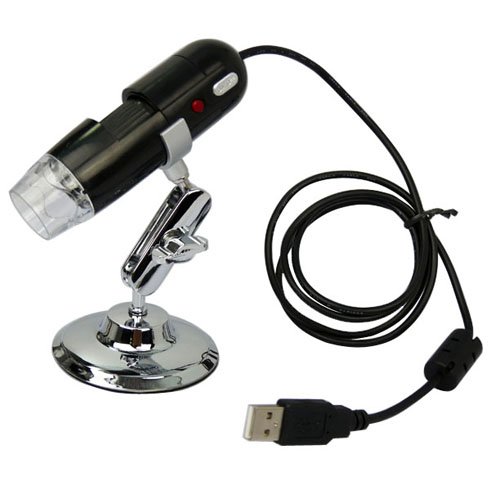
GSI High-Definition Scientific Digital LED Microscope, USB Video Connection to Computers/Laptops/Notebooks, Magnifies All Microscopic Articles 200x, Special Image Treatment, Includes Stand for Handsfree Use, for Hobby/Science/Education/Industrial Review


GSI High-Definition Scientific Digital LED Microscope, USB Video Connection to Computers/Laptops/Notebooks, Magnifies All Microscopic Articles 200x, Special Image Treatment, Includes Stand for Handsfree Use, for Hobby/Science/Education/Industrial Feature
- Microscope Lens (25x - 200x), 4 LED Light Source, Snap Function
- Color CMOS Image Censor, High Speed CPU, 1280x960 Resolution, 1.3M Pixels
- USB Compatible for Instant Video Viewing on Computer or Laptop Screen
- Auto White Balance Control, Sharpness Control, Color Mangement, Brightness Adjustable
- High Quality Image Enlarges Microscopic Articles on Skin, Cloth, Leather, Coins, Bills and Hair, Etc.
GSI High-Definition Scientific Digital LED Microscope, USB Video Connection to Computers/Laptops/Notebooks, Magnifies All Microscopic Articles 200x, Special Image Treatment, Includes Stand for Handsfree Use, for Hobby/Science/Education/Industrial Overview
This season discover the Micro world with this newest innovation from GSI. This product was designed by our World-Class Micro experts to capture and display the tiniest miniscule objects in 200 times magnified viewing. Connect to your computer with USB cable included, simply point-and-Snap, and watch the world come alive in front of your eyes.Related Products
- Carson MM-200 Carson Micromax LED 60X-100X LED Lighted Pocket Microscope
- Celestron 44302 Deluxe Handheld Digital Microscope 2MP
- SE MW10088UV Pocket Microscope with UV Light
- SE MW10087L Mini Brass Microscope with Illuminator
- SE 30X Illuminated Jewelers Loupe
Customer Reviews
*** Product Information and Prices Stored: Oct 11, 2012 01:45:05
My Links : Best Fasteners Measurement Guide droid cell phone covers