Wednesday, October 31, 2012

Reviewing the Types of Light Microscopes

A light microscope is one of the most beloved types of microscopes for finding at images. In the beginning, light scopes were created to see things such as fabric weave and other uncomplicated objects.

However, as time progressed, a person named Anton van Leeuwenhoek found ways to create scopes that allowed population to see things that were previously invisible. Today, these microscopes have come a long way but they still use light in assorted ways to view "invisible" worlds.

Light Microscope Images

One of the most beloved types of a light microscope is that of the captivating field microscope. These are the most beloved in most schools. A captivating field microscope aims light straight through the specimen into the eyepiece, in order to view the objects on the microscope slides.

A student can then use the coarse or fine knobs to adjust their field of view. Unfortunately, these are microscopic light microscopes and can sometimes distort the image as soon as one begins to see it clearly.

Another type of light microscopes that can be found in many labs is that of the dark field. These microscopes are great for viewing objects that would otherwise seem distorted with a captivating field scope, such as cells and pond water.

The small metazoans can for real be seen and remarkable when using this type of scope. The light is reflected by particles on the microscope slide. This allows for a more in depth study at a small cost compared to a scanning electron one.

Choosing a light microscope to look at items can be a daunting task. One has basically two choices and depending on how much or what one wishes to see will conclude if a person chooses correctly. Both the dark and captivating field microscopes offer great advantages for those who want to see the unseen.

However, if one wants to be more in depth, then a microscopic more money and effort studying about the in's and outs of the scope will be required. The main thing to remember is that science is the backbone of our world, no matter how high tech it has become.

Reviewing the Types of Light Microscopes

Friends Link : Pneumatics and Plumbing Good choice Men Eco Drive Watch Cell Phone Backup

Tuesday, October 30, 2012

BigC Dino-Lite Digital Microscope AM313 with measurement function


BigC Dino-Lite Digital Microscope AM313 with measurement function Review


BigC Dino-Lite Digital Microscope AM313 with measurement function Feature

  • *Stands (MS22B) are sold seperately!
  • Resolution: 640* 480pixels (VGA)
  • Object measurement function by software
  • Adjustable magnification: 10x, 20x~200x (Continuous magnification without switch lenses)
  • Built-in white-light 8 LED illumination

BigC Dino-Lite Digital Microscope AM313 with measurement function Overview

** Pictures shown with optional MS22B Stand which can be purchased seperately!Dino-Lite PLUS Digital Microscope with USB output (AM313)Easy and convenient to operate! 10x ~ 200x LED on/off and Measurement object feature controlled by softwareDino digital microscope provides high quality microscopy video interfacing to PC. Clear and steady imaging with simple magnification and focus control.Resolution: 640* 480pixels (VGA) Adjustable magnification: 10x, 20x~200x (Continuous magnification without switch lenses) Built-in white-light 8 LED illumination Frame rate: up to 30fps Interface: USB (PC) Dimension: 10.2cm (Height) X 3.2cm (D) Weight: 95g With Dino Capture SoftwareEnabling you to capture image, video and time-lapsed video in a click.
*** Product Information and Prices Stored: Oct 30, 2012 09:15:03

See Also : The Fasteners Finishing Products Pneumatic Plumbing the compound microscope samsung batteries for cell phones

Monday, October 29, 2012

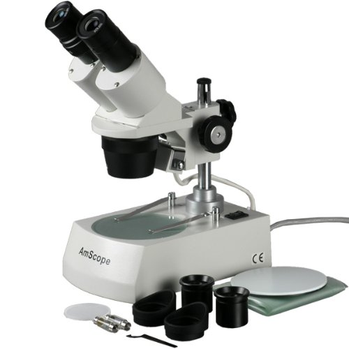
AmScope New! Dissecting Binocular Stereo Microscope 20x 40x 80x

AmScope New! Dissecting Binocular Stereo Microscope 20x 40x 80x Review


Prices as of : Oct 29, 2012 02:00:04
Listprice : $700.00
Offier Price : $138.75
You Save : $561.25

AmScope New! Dissecting Binocular Stereo Microscope 20x 40x 80x Feature

  • Three Magnification Power Settings: 20X, 40X & 80X
  • Built-In Both Incident (top) Lighting and Transmitted (bottom) Lighting
  • Sharp Stereo Erect Images over a Wide Field View
  • Great for Exploration of the World of Surface Details and Fine Structure
  • Perfect for Examination of Insects, Plants, Rocks, Stamps, Coins, Jewel, Soldered Points, Microelectronic Components or Virtually any 3-D Objects

AmScope New! Dissecting Binocular Stereo Microscope 20x 40x 80x Overview

This binocular stereo microscope comes with three magnification power settings (20X, 40X & 80X) and a versatile illumination system that provides both incident (top) lighting and transmitted (bottom) lighting. You can choose between incident illumination shining down onto the object or transmitted illumination through the frosted stage plate. The first is used for the observation of three-dimensional objects and the second for the observation of slides. This microscope offers high resolution and good depth within a broad field of view. It gives sharp clear stereo images. Its 45° inclined binocular head ensures an easy observation and rubber eyeguards provide further comfort. This microscope is useful for many applications. It can be used in geology, forestry, archaeology, agriculture and biology. It is appropriate for educational purposes in hospitals and schools. It is great for the exploration of the world of surface details and fine structures of various materials like wood, metal, solder, semiconductor and stone. It is perfect for the examination of insects, plants, rocks, stamps, coins, jewel, soldered points, microelectronic components or virtually any three dimensional objects. This scope is an ideal tool for watchmaker, jeweler, engraver, dies-maker, philatelist, coin collector, students, and etc. It comes with first-class quality and affordable price. It is brand new in factory-sealed box. The retail value of this microscope is 0.

*** Product Information and Prices Stored: Oct 29, 2012 02:00:04

See Also : Picking Safety Products Sun Solar cell Measurement Guide led microscope light

Saturday, October 27, 2012

7th Grade Science project Ideas Made easy and Fun

There are a whole of 7th grade science scheme ideas that a pupil can select from to demonstrate concepts learned in class. A few such projects include:

o derive water from a colse to stream or pond and view a drop of it under a microscope. Note what kinds of things you see and make drawings of them. Which are plants and which are animals? What is greater in your sample, plants or animals? What factors might affect the numbers of each that you find?

Light Microscope Images

o Draw a cell and label all of its parts. What function does each part of the cell serve? Are there differences in the middle of plant and animal cells? If do, what are they?

o Name the four separate blood groups found in humans. What type is the rarest? The most common? How does the blood type of a person's parents affect the outcome of their own (i.e. If a person with type A blood marries person with type O, what type will any offspring have)?

o Draw a photo of a red blood call from a human and label all of its parts. Now draw a photo of the red blood cell from an animal, like a cow and label all of the parts. Are there any differences? If so, what part do you think these differences play in the function of the cell?

o Build a 3D model of a single-celled animal from cake. Use separate tints of frosting to make the separate structures inside the animal. Licorice laces can be cut up to recount the cilia. Before the cake is eaten, hand out a sheet that has the separate parts labeled with call-outs, but not filled in. Allow population to view the cake and fill in the papers as to what each buildings is. Just before the cake is cut, put out a poster that shows what everything is. Let the person who has the most right answers for these 7th grade science scheme ideas have the first piece of cake.

o Take a sample of cells from the inside of the mouth and view them under a microscope. Stain the material with a drop of food coloring. Recite what you see. Make a drawing of any clear cells and label all of the structures present.

o Plankton is made up of several kinds of particular cell plants and animals. Find out what they are and make drawings of several separate kinds of each. What animals use plankton as a food source. What kinds of changes in the environment could change plankton's survival in the water? Has plankton levels risen or fallen in new years? Why?

o separate bacteria exist all colse to us. Where are they found? What role do they play? In what cases should bacteria be encouraged to grow? In what cases should they be discouraged? Give an example of how or why they should be encouraged, and why they should be discouraged.

With a little bit of rigorous opinion it is possible to find any whole of 7th grade science scheme ideas that can be turned into a science scheme for the inquisitive student.

7th Grade Science project Ideas Made easy and Fun

Thanks To : Measurement Guide Finishing Products kodack easy share

Thursday, October 25, 2012

Light Microscopes

Light microscopes or enthralling field microscopes are the most basic form of microscopes. The customary light microscope was invented way back in the 16th century. They are so called because they use light in the descriptive spectrum to illuminate the specimen. Light microscopes are extensively used in any place from high-tech laboratories to schools.

There are many types of light microscopes ready from just to 00 or even more. The major types of light microscopes are stereo or dissecting, simple and blend microscopes. Stereo microscopes are the most advanced and use two light paths for three-dimensional (3D) viewing. They are very expensive and are used in laboratories and corrective practices. simple light microscopes are used only for small scale magnification and they have only one lens both as eyepiece and objective.

Light Microscope Images

Compound microscopes are the most extensively used light microscopes. There are mainly three grades of blend microscopes - student, benchtop and explore light microscopes. Pupil microscopes are cheap, small and designed for enthralling field, dark field, and phase difference examinations. Benchtop microscopes are used for test techniques. explore microscopes are for explore practices; they often comprise built-in computers and cameras.

The components of a coarse blend microscope are: a mirror or light source for reflecting or producing light, a condenser with diaphragms and filters to adjust the intensity of the light, a stage for supporting the specimen which contains a central hole to pass the light on to the specimen, variable objective lens for focusing the image for the eyepiece, fixed eyepiece or ocular for focusing the image for the eye, screws known as adjustments (coarse adjustment and fine adjustment) are used to bring the specimen in to focus, and a framework to which all these parts are attached. The power of the microscope depends on the power of the objective and eyepiece.

If you intend to buy a light microscope, go to specialist microscope suppliers, because only they can give you satisfying after-purchase services. The Internet also provides data about some favorite suppliers. Often Internet sites offer you good deals and services.

Light Microscopes

Recommend : Measurement Guide Fasteners Equipment Pneumatics and Plumbing Good choice cell phone plans for kids blu ray player hd wifi

Wednesday, October 24, 2012

Microphotography - The Art and Science of Taking Pictures straight through a Microscope

Taking digital photos through a microscope is the best way to sustain the images that you observed through microscope lens. The digital photos can be used for documentation, publication or just for fun of sharing the wonderful view you have. It can be entertaining because of the low light conditions. However, taking pictures through microscope have never been easier with the advances in digital technology. In this article, I want to introduce you four different ways to take photos through a microscope.

Taking pictures with snapshot cameras: Most buyer cameras are snapshot cameras. The ability of the cameras in this class is enhancing and the prices of these cameras are continued to drop. (An entry level Canon powershot® camera, beginning nearby 0s at the time of writing). These cameras also come with many pre-configured settings suitable for different conditions. Taking photos with snapshot cameras is relatively fool proof. Although convenient, none of the preset configuration is optimized for microphotography. You typically need to adjust the camera zoom, white balance, exposure recompense to optimize the photo quality.

Light Microscope Images

How To:To take the pictures using snapshot camera, you need to get the camera as close to the eyepiece as inherent then take picture directly through the eyepiece. The main challenge of this formula is the mystery of locating ideal placement of the camera and to hold it still. You can indubitably fix the problem by attaching a commercially available universal camera adapter to the tripod socket then attach the adapter to the microscope eyepiece.

Taking pictures with Dslr cameras: Dslr cameras are mostly used by professional and hobbyists to capture professional photographs. The picture ability is first-rate to most snapshot cameras. They usually allow you to use interchangeable lens. The ability to configure the manual setting also is also first-rate to snapshot cameras.. You may set the extended exposure time or high sensitivity to optimize the photo shooting at low light condition.

How to: To take the pictures with the Dslr through a microscope, remove the lens and attach the camera to the microscope camera adapter which essentially is a camera adapter with build-in eyepiece. Some Dslr models offer the remote control and previewed on computer screen which is much larger than the small view finder of the camera.

Microscope camera: These cameras are specifically designed for microscopes. They typically cost more for less resolution, but they have the advantage of directly controlled by computers. Images and videos can be captured to the computer hard drive without having to change the images from the flash disk. The qualities of these cameras varied greatly. The resolutions of these cameras range from 640 x 480 to several mega pixels. There are two types of chips inside microscope cameras - the Ccd (charged coupled device) chip and the Cmos (complementary metal oxide semiconductor) chip. A Ccd chip generally produces higher ability images in low-light conditions and is better for publication ability images. Cameras with Ccd chips generally cost more than those with Cmos chips. Unlike the Dslr or snap shot cameras which have many buttons. These cameras typically controlled by computers using Usb connection.

How to: To take photos with microscope cameras, you need to install the camera driver and image capture software. These cameras can be attached to a microscope adapter like the Dslr and drop into the eyepiece socket for photo taking. The configurations of cameras are usually controlled by computer.

Build-in Digital Camera: Some microscopes come with a build-in camera. These cameras are in fact a microscope cameras attached to a third port of a trinocular microscope. The photo ability of these cameras is usually better than others - not in terms of mega pixel, but with better resolution, field of view and integration.

How to: Taking photos with microscope build-in digital camera is painless. You can whether taking the photos with the camera shooter button or with image capturing software. No adapter is needed. However, they usually cost more than other options.

Microphotography - The Art and Science of Taking Pictures straight through a Microscope

Tags : Finishing Products Pneumatic Plumbing light microscope image

Tuesday, October 23, 2012

theory of Microscopes and Microscopy - Part I

The microscope is possibly the most tasteless tool in any microbiology lab, since it enables students and professionals to contemplate organisms and structures invisibles to the naked eye. There are microscopes with some dissimilar magnification levels, fluctuating from hundreds to thousands of hundreds in diameter. Of course, the price of a microscope increases with its quality, but even the simplest microscope can supply breathtaking views of the microbiology world.

Each kind of microscope and each technique of material making ready offers some exact advantages on the demonstration of inevitable morphological elements. In this description series, we will learn the ideas and types of microscopes, as well as some of the microbiological methods used to contemplate the dimensions, the shapes and structural characteristics of microorganisms.

Light Microscope Images

The microscopes fit into two categories: light (or optical) and electronic, depending on the principle or method of magnification employed. In light microscopy, the magnification is obtained straight through a ideas of optic lenses while in electronic microscopy a bean of electrons is used to yield the enlarged image.

The light microscopy can be divided into six sub-categories:

Bright field microscopy Dark field optics Ultraviolet microscopy Fluorescence microscopy Phase incompatibility microscopy Differential interference incompatibility (D.I.C).

Beginners will much probably use entertaining field microscopy to contemplate their samples, since it is by far the most tasteless technique employed in explore and quality operate labs. However, it is very foremost to know the other applications, for each one has a unique asset that makes it especially desirable for the demonstration of exact morphological structures.

See you tomorrow for the next article: parts and functions of a light microscope.

theory of Microscopes and Microscopy - Part I

Visit : Best Fasteners Sun Solar cell Finishing Products best hard disk media player

Monday, October 22, 2012

AmScope 7X-45X Stereo Zoom Boom Microscope + Ring Light

AmScope 7X-45X Stereo Zoom Boom Microscope + Ring Light Review


$1231

AmScope 7X-45X Stereo Zoom Boom Microscope + Ring Light Feature

  • 7X-45X large zoom magnification power on flexible boom stand
  • Crystal clear original true color images
  • Fluorescent Ring Light Included
  • 4" (100mm) Extremely Large Working Distance
  • Excellent five (5) year manufacturer warranty

AmScope 7X-45X Stereo Zoom Boom Microscope + Ring Light Overview

This is a binocular 7-45X zoom stereo microscope on boom with fluorescent ring light . It comes with a 45° inclined binocular head, super widefield high-eyepoint eyepieces, a heavy-duty boom stand and a fluorescent ring light. The 45° inclined eye tube and high-eyepoint optic design ensure an easy and comfortable observation, especially for eyeglass wearers. The rubber eyeguards provide further comfort. The microscope offers high resolution, super widefield of view, large magnifying zoom range and long working distance. It gives crystal clear sharp images. The fluorescent ring light provides cool, even and bright illumination. It is an ideal instrument for research labs, gemologists, engravers, collectors and anyone in semiconductor and/or electronic industry. This microscope is made by the same technicians and on the same production line as optical instruments for Leica, Zeiss, Nikon and Olympus. It is brand new in factory box. The retail value of this microscope is ,600.

*** Product Information and Prices Stored: Oct 22, 2012 02:45:06
$369.00

My Links : Measurement Guide Best Fasteners Measurement Guide Digital Camera

Sunday, October 21, 2012

What Are The distinct Kinds Of Microscope?

Microscopes are one of the most leading inventions of all time. They have helped the lives of every population to become best and easier. They have helped population in the fields of biology, astronomy, geology, medicine, technology, and approximately in everything. There are many dissimilar kinds of microscopes

today, each with dissimilar use. This article will justify each type.

Light Microscope Images

1. Combination Microscope - These are the ones that are commonly seen and used everywhere. They are the ones that you see and use in most biology and other science classes. Some are electrically operated. Combination microscope uses light to heighten the image of the subject a man is viewing. They have complicated lenses for viewing in dissimilar magnification. 4x, 10, 40x, 100x are the most tasteless magnifying power of this device, excluding the eyepiece.

2. Dissecting or Stereo Microscopes - These have low magnification power and, like Combination microscopes, they are also light-powered. Compared to a Combination microscope though, they have a higher magnification and are also able to deliver a 3 dimension view of the subject being viewed at.

3. Scanning Electron Microscope or Sem - It is a type of microscope that is very dissimilar from Combination and dissecting microscopes because instead of using light to produce an image they use electrons to do this. Just like dissecting microscope, they also produce images in 3 dimensions except that they produce these images in a much higher resolution and best magnification. Sem's also have a best depth of focus. Scanning electron microscopes are used in many things these days together with science research, medicine, and forensics.

4. Transmission Electron Microscope or Tem - Transmission electron microscopes are microscopes that uses electrons instead of light to produce an image. The subject that is to be viewed should be very thin for an image to be produced. Tems are able to produce high resolution and magnification images. Unlike Sem's, which produce 3 dimension images, Tem's are only able to give two dimension images.

Being able to know the dissimilar kinds of microscope and their uses is a major benefit for you especially if you are a student. The advancement of technology has enabled us to construct newer and best instruments which let us see things that we have not seen before using our naked eye.

Knowing the dissimilar ways we can use scientific instruments can be a boost to our knowledge. When we know how to use these instruments, we then have an opening to widen our learning.

What Are The distinct Kinds Of Microscope?

Visit : Pneumatics and Plumbing Good choice Finishing Products blu ray player hd wifi

Friday, October 19, 2012

Aven 26700-206 112mm Working Distance Digital Microscope with Polarizer USB, 10x - 200x Magnification

Aven 26700-206 112mm Working Distance Digital Microscope with Polarizer USB, 10x - 200x Magnification Review


Prices as of : Oct 19, 2012 10:00:03
Listprice : $262.11
Offier Price : $178.75
You Save : $83.36

Aven 26700-206 112mm Working Distance Digital Microscope with Polarizer USB, 10x - 200x Magnification Feature

  • Digital, hand-held microscope with 1/4" color complementary metal-oxide semiconductor (CMOS) image sensor, polarizer, and USB 2.0 computer output to display images, for inspection of reflective surfaces, measurement, and analysis
  • Optical magnification of 10X to 200X, and resolution output of 1280 x 1024 National Television System Committee (NTSC) standards
  • Six white LED lights with adjustable brightness to control illumination, automatic white balance, and the ability to be turned on and off
  • Included software captures images and has measurement and image comparison functions.
  • One year warranty

Aven 26700-206 112mm Working Distance Digital Microscope with Polarizer USB, 10x - 200x Magnification Overview

The Aven 26700-206 Mighty Scope with polarizer is a hand-held, digital microscope that has a 2.0 USB interface, six white LED lights, a Microtouch shutter trigger, optical magnification ability of 10X to 200X, and comes with image and video capture software. This digital microscope enhances the resolution of an object or image, and connects to a computer for image display. It is commonly used for quality control (QC) inspection, medical and scientific research, geology, mineralogy, chemistry, and for printing and textile inspection.

This mobile microscope houses a polarizer to inspect reflective surfaces, and produce clear images. Its adjustable magnification of 10X to 200X comes in an all-in-one, compact lens. A 1/4” complementary metal-oxide semiconductor (CMOS) color image sensor manages noise reduction and image quality, and delivers high-resolution, 1280 x 1024 National Television System Committee (NTSC)-standard pixel images. The frame rate is 30 frames per second (FPS), and 15 FPS at 1.3 mega-pixels. The Mighty Scope uses six white LED lights with adjustable brightness to control illumination. Microscope lighting can be turned on and off. The microscope has automatic white balance to control brightness, and automatic gain control to amplify the electrical output and show a clearer image. A Microtouch shutter trigger allows for convenient image capture when in hand-held mode, or mounted on a stand. The microscope has a 112 mm working distance. Included software captures images and has measurement and image comparison functions. The microscope comes with an adjustable metal stand that includes height and angle adjustment. The Mighty Scope with polarizer microscope has a one-year warranty.

Specifications

Optical magnification10X to 200X
Image sensor0.25” color CMOS
Effective pixels 1.3 mega-pixels (M)
Illumination6 adjustable, white LED lights
Gain controlAutomatic
White balanceAutomatic
Snap shot modeHardware and software controllable
Working distance (WD)10X at 112 mm, 20X at 72mm, 30X at 19mm, 40X at 8.5 mm
Frame rate30fps, 15 fps at 1.3 M
Interpupillary adjustment52 to 75 mm
Operating systemWindows XP, SP2, and Vista 7
Power source5 VDC through 2.0 USB port
Power consumption110-volt
Overall dimensions22.9 x 12.7 x 15.9 cm / 9 x 5 x 6.25 inches (H x W x D)
Weight0.703068 kilograms (1.55 lb.)
WarrantyOne year

*(H is height, the vertical distance from the lowest to highest point; W is width, the horizontal distance from left to right; D is depth, the horizontal distance from front to back.)

Microscopes are instruments used to enhance the resolution of an object or image. Types include compound, stereo, or digital. Compound microscopes use a compound optical system with an objective lens and an eyepiece. Stereo microscopes show object depth in a three-dimensional image. Digital microscopes are used to display an image on a monitor, rather than looking through a lens. Microscopes can have monocular (one), binocular (two), or trinocular (three) eyepieces, with varying magnification abilities. Magnification ability refers to the size of an image. Resolution, also known as resolvant power, refers to the clarity of the image. The interaction between field of view (FOV), numerical aperture (NA), and working distance (WD) determines resolution. Microscopes can control magnification through a fixed focus, or through a range of adjustments. They can also utilize LED, fluorescent, and mirror light sources to help control viewing capabilities. Microscopes are widely used in education, lab research, biology, metallurgy, engineering, chemistry, manufacturing, and in the medical, forensic science, and veterinary industries.

Aven manufactures optical inspection tools, precision tools, and bench accessories for industrial, scientific, and research applications. The company was founded in 1983 and is headquartered in Ann Arbor, MI.

What’s in the Box?

  • 26700-206 Mighty Scope microscope
  • Adjustable metal stand
  • Instructions


*** Product Information and Prices Stored: Oct 19, 2012 10:00:03

My Links : The Pneumatic Fasteners Equipment The Fasteners blu ray player hd wifi

Thursday, October 18, 2012

Types of composition optic Microscopes

Compound optic microscopes are also known as composition microscopes, light microscopes or optic microscopes and come in some varieties. These comprise digital, inverted, stereo, monocular and binocular.

All work on the same system and deliver an enlarged image to the viewer. They have some differences though, which will be detailed here.

Light Microscope Images

Monocular:

These are probably the ones you think of when microscopes come to mind. Using a particular light as a sample illuminator and composition lenses for magnification, these have a particular eyepiece. This is a bit uncomfortable, as you need to close one eye to get a clear image of what you are looking at on the slide. The eyepiece has a power of about 10X and the objectives or lenses on the nosepiece range from 2X to 50X depending on your particular microscope. The big one here is the particular eyepiece.

Binocular:

Binocular microscopes are becoming more common. You can tell a binocular style microscope by the dual eyepiece. Simply put, you use both eyes to view the sample image. This makes them more comfortable to use and thereby more popular. Dual eyepiece scopes are used widely in high school and college classrooms. Binocular microscopes have all the same characteristics as the monocular ones above.

Stereo:

Stereo microscopes bring a whole new dimension to the image, literally. Whereas the typical light microscope produces a two-dimensional image, the stereo microscope uses two light sources working independently to yield a three-dimensional image to the viewer. The sample on the slide will have height, depth and width. Using all of the same features and characteristics of the other optic microscopes, stereomicroscopes stand out among their counterparts.

Digital:

This is where things go differently in the realms of microscopy. Thorough optic microscopes use light to illuminate and lenses to magnify something that you could not see with the unaided eye.

Digital microscopes are a breed apart. This type of microscope inverts the light source, placing it above the sample. Thorough optic microscopes place the light below the sample. You also get the 3D image like in stereo scopes. The discrepancy is that the image is digitized and transmitted to a monitor or screen for viewing. Dream watching cells divide on a 19" monitor. The user can take still photos or consuming video of the sample in real time. I think you can see the advantages here.

Inverted:

Inverted microscopes are used to study samples that are gravity sensitive, like gases suspended in a liquid. The inversion refers to the light source, which is typically below the sample slide. With the inverted microscope, the light source is above the sample slide. This paved the way for new innovations in digital imaging that came along.

All these distinct types will work for you. It's just a matter of what you want to achieve or study. I can tell you this, though; using any microscope will open your eyes to a whole new perspective of where we came from and what we are made of.

Types of composition optic Microscopes

Related : Fasteners Equipment best unlimited cell phone plan

Wednesday, October 17, 2012

AmScope 40X-1000X All-Metal Cordless LED Student Compound Microscope with Coarse & Fine Focus + Digital Imager

AmScope 40X-1000X All-Metal Cordless LED Student Compound Microscope with Coarse & Fine Focus + Digital Imager Review


Prices as of : Oct 17, 2012 04:00:04
Listprice : $500.00
Offier Price : $119.98
You Save : $380.02

AmScope 40X-1000X All-Metal Cordless LED Student Compound Microscope with Coarse & Fine Focus + Digital Imager Feature

  • 360° Rotatable Monocular Offering Five Magnification Settings up to 1000X
  • View and/or Capture Snapshot Images or Video with Your Computer; User-friendly Software Compatible with Windows XP/Vista/7
  • Co-axial Coarse & Fine Focus, Single Lens Condenser and Disc Diaphragm
  • LED Illumination by Wall-power and/or Batteries (wall-power adapter/recharger included)
  • Sturdy All-Metal Framework

AmScope 40X-1000X All-Metal Cordless LED Student Compound Microscope with Coarse & Fine Focus + Digital Imager Overview

This microscope with a digital imager (digital camera) is designed for students to learn science, offering five magnification settings, 40X, 100X, 250X, 400X and 1000X. It comes with a 640x480 pixel USB2.0 imager, full optical glass elements, all metal framework, 360° rotatable monocular head, coarse & fine focus, and the LED illumination system that uses either wall-power (adapter included) or three AA batteries (or re-chargeable batteries). The camera can replace the microscope eyepiece so you can view comfortably on your computer screen. CD-ROM contains software for viewing and image capture when using the imager, and works with Windows XP, Vista, and 7 operating systems. It is an ideal microscope for home school uses or for students from elementary to high school. It is also an excellent instrument for hobbyists. It is brand new in original box. Its retail value is 0.

*** Product Information and Prices Stored: Oct 17, 2012 04:00:04

My Links : Pneumatics and Plumbing Good choice Measurement Guide Sun Solar cell light microscope magnification games for cell phones

Monday, October 15, 2012

How Each of the Microscope Parts Functions

Understanding how each of the microscope parts functions is prominent if you're going to truly understand the microscope. This will make you more of an devotee on the field and allow you to intelligently evaluate how well one is working--and either it needs a exchange part. With that in mind, let's scrutinize the field of the microscope's parts and how they work.

Generations of habitancy nearby the world have made use of the microscope. Straight through the decades and centuries, its basic establish has remained much the same. Even though microscopes have changed Straight through the years, from the basic light much microscope all the way to electron microscopes, the parts and functions of a compound microscope are remarkably the same.

Microscope

A microscope will only work correctly as long as each of its private parts is working correctly. When one part ceases working properly, it will likely hinder the effectiveness of the instrument altogether. The main parts of most microscopes are the tube, lenses, illuminator, arm, adjustment knobs, and stage.

You'll find two basic kinds of lenses on the typical microscope. First of the lenses is the eyepiece lens (or the "ocular lens," as it's also called). This ocular lens is found at the microscope's top. This is the part that the microscope user looks through. It's typically not adjustable. The microscope's second lens is known as the objective lens. It's the one that provides most of the instrument's magnification. Indeed, most microscopes don't have one, but some objective lenses. Each objective lens varies in magnification strength.

The microscope's objective lenses are part of a circular part of the scope. It's found in the middle of the eyepiece and stage. The user selects the objective lens based on the power that he needs and the power provided by that objective lens. If the user desires a separate zoom level, he rotates the circular disc, thus placing a separate lens above the stage. Connecting the ocular lens and the objective lenses are the part of the microscope called the tube. The user looks Straight through the ocular lens and Straight through the tube, ultimately finding out of the bottom, Straight through one of the objective lenses.

The specimen or object to be examined is settled on a part called the stage. Slides are secured to the stage by use of clamps. On these slides will be the specimens to be examined--specimens such as blood or micro-organisms. Immediately below the stage is something such as a mirror or, on a compound light microscope, a light. This mirror or light is called the illuminator, and it's what makes it easier for the user to see the specimen.

Finally, there is a pair of adjustment knobs on most types of microscope. The adjusters are used to support in focusing the lenses. The coarse one is the larger of these two knobs--the one that pulls the lens and stage nearer together. The fine adjuster is the smaller of the adjustment knobs. First, the user adjusts the coarse knob and then the smaller one to give the tiny adjustments needed to bring the object into clearer focus.

These microscope parts and functions are the same on almost all microscopes that you'll encounter. There is some small variation, with slightly separate parts on some (For example, on an electron microscope, there will be electron beams rather than typical illuminators; it thus varies a bit from the parts and functions of the compound microscope). Yet the basic functions and parts are the same. If you learn how each of these microscope parts functions, it will be easier for you to know a good microscope from a bad one.

How Each of the Microscope Parts Functions

Thanks To : Picking Safety Products Pneumatics and Plumbing Good choice microscope light

Sunday, October 14, 2012

What Are the Types of Microscopes?

An electron microscope can offer a person many separate types of advantages over other microscopes. If person has never used one of the many types of electron microscopes, then they are seriously missing out.

This type of microscope offers more clarity and a great magnification than most any other type of microscope on the market. In order to select the best type of the many electron microscopes, a person must understand the various types that are ready for them to enjoy.

Microscope

Three types of them exist. They are the transmission electron microscope, the scanning electron microscope, and the scanning-tunneling electron microscope. The primary of all of these types of devices is the transmission electron.

The Tem, as it is ordinarily called, transmits electrons through a small slice of a specimen in order for the image to be seen. These types of devices are great for looking inside the cells of beings on the planet. Much has been learned by using these microscopes for looking all types of various cells.

Other types of the electron expedient are the scanning tunneling microscope and the scanning one. Both types of devices allow for a more in depth look than most other type of microscopes. The scanning microscope can well allow scientists to view items in 3-D because of the excited atoms that are on a specimen.

The tunneling microscope well creates a tunneling current between the electrons on a specimen and the microscopes tip to make the image. These are both very high tech microscopes that can be used to see farther into a specimen than previously view possible.

An electron one can give a much more in depth view. Population once only had an idea of what cells and other imperceptible items looked like. Thanks to the invention of the electron microscopes and the various types that are available, a person can see these items in a three dimensional view that can make learning these items better. Microscopes are the wave of the future in today's current high tech world.

What Are the Types of Microscopes?

See Also : Picking Safety Products The Pneumatic Measurement Guide cheap family cell phone plans best unlimited cell phone plan

Saturday, October 13, 2012

The development of the Microscope

An object that is observed under microscope can be immortalized by using camera. A blend microscope that has two ocular lenses is ordinarily completed with the lens part for camera.

Technology as the work consequent of human all the time experiences the developments. Easy microscope and some other visual microscopes are only able to improve the object of about 100-1000 times, whereas electron microscope technology can be enlarged up to 1 million times.

Microscope

Based on the illumination system, this tool is divided into two, namely visual and non-optical microscope.

a. visual microscope

Its magnification process of objects uses general light (visible light). It includes compound, binocular, and also stereoscopy binocular microscope which produces the image of 3 dimensions, and ultraviolet microscopy.

b. Non-optical microscope

It amplifies the objects by being helped by the short wavelength radiation. It includes X-ray, ion, and electron microscope. Among the three types of non-optical microscope, the most widely used is electron microscope.

Through the electron one, you can learn the patterns of animal cells,
plants, and bacteria. It is also used in analyzing the consequent of business and controlling the production result.

The electron microscope is a microscope that is able to perform the magnification of objects up to 2 million times. It uses electro-static and electro magnetic to control the lighting and the display of images. It also has the zoom quality and resolution that is much good than light microscopy. In this case, it uses more power and shorter electromagnetic radiation than the light microscope.

The development of the Microscope

Related : The Fasteners Measurement Guide Finishing Products games for cell phones

Friday, October 12, 2012

iOptron ST-640 Digital Microscope with LCD Screen

iOptron ST-640 Digital Microscope with LCD Screen Review


Prices as of : Oct 12, 2012 07:45:03
Listprice : $308.00
Offier Price : $186.11
You Save : $121.89

iOptron ST-640 Digital Microscope with LCD Screen Feature

  • LCD Module with 3.5" LCD Screen, photo and video capturing capabilities, 8x zoom
  • 10x, 16x Wide Field Eyepiece. 4x, 10x, 40x Objectives.
  • 640x maximum magnification
  • 2 LED illumination light sources for brighter, longer and energy-saving usage.
  • Save video and photos to your computer!

iOptron ST-640 Digital Microscope with LCD Screen Overview

This high-tech microscope is capable of both digital viewing through a large 3.5" LCD screen and via traditional eyepieces for additional magnification. The camera imbedded in the digital eyepiece can shoot both pictures and videos. By simply connecting the microscope to your computer you can easily browse and organize pictures and videos. The ST-640 LCD Microscope makes studying plant and insect samples easy and fun for the whole family.

*** Product Information and Prices Stored: Oct 12, 2012 07:45:03

Thanks To : Fasteners Equipment Measurement Guide Pneumatic Plumbing samsung cell phone charger underwater canon digital camera

Wednesday, October 10, 2012

Great Price GSI GWC60-1 for $33.49


GSI High-Definition Scientific Digital LED Microscope, USB Video Connection to Computers/Laptops/Notebooks, Magnifies All Microscopic Articles 200x, Special Image Treatment, Includes Stand for Handsfree Use, for Hobby/Science/Education/Industrial Review


GSI High-Definition Scientific Digital LED Microscope, USB Video Connection to Computers/Laptops/Notebooks, Magnifies All Microscopic Articles 200x, Special Image Treatment, Includes Stand for Handsfree Use, for Hobby/Science/Education/Industrial Feature

  • Microscope Lens (25x - 200x), 4 LED Light Source, Snap Function
  • Color CMOS Image Censor, High Speed CPU, 1280x960 Resolution, 1.3M Pixels
  • USB Compatible for Instant Video Viewing on Computer or Laptop Screen
  • Auto White Balance Control, Sharpness Control, Color Mangement, Brightness Adjustable
  • High Quality Image Enlarges Microscopic Articles on Skin, Cloth, Leather, Coins, Bills and Hair, Etc.

GSI High-Definition Scientific Digital LED Microscope, USB Video Connection to Computers/Laptops/Notebooks, Magnifies All Microscopic Articles 200x, Special Image Treatment, Includes Stand for Handsfree Use, for Hobby/Science/Education/Industrial Overview

This season discover the Micro world with this newest innovation from GSI. This product was designed by our World-Class Micro experts to capture and display the tiniest miniscule objects in 200 times magnified viewing. Connect to your computer with USB cable included, simply point-and-Snap, and watch the world come alive in front of your eyes.

Related Products

Customer Reviews


*** Product Information and Prices Stored: Oct 11, 2012 01:45:05

My Links : Best Fasteners Measurement Guide droid cell phone covers

Tuesday, October 9, 2012

The Electron Microscope Does Have Disadvantages

The world has come a long way from the first light microscopes that were used, and one type of microscope for study that is very beloved is that of the electron microscopes. Researchers all over the world use these extremely technical scopes on a daily basis to find worlds that were unheard of until today's high tech world made them possible. However, issues plague these microscopes for study purposes, and one should without fail take the time to rejoinder a few questions before they spend in one.

The first major issue with such a microscope is that it is very expensive. The presume for this is naturally because of the voltage that is required in order to control these microscopes.

Light Microscope Images

Light microscopy and the microscopes that are used with it are great for the average hobbyist, but the electron microscope is not for those people. These expensive magnifying tools can run into the thousands of dollars for the first cost and the electricity bill will be outrageous in the end.

Another major disadvantage of the electron microscopes is that the microscope images have to be viewed in a vacuum. The specimens also need wide establishment in order for them to be looked at under this microscope.

However, the images are astonishing and can be viewed in a three dimensional image with a scanning electron microscope. The environmental scanning one is the only irregularity in the electron microscope class that does not have to view the samples in a vacuum. These images should be seen in a low pressure, wet environment.

Electron microscopes can prove to be a huge asset to many study labs over the world. However, if one does not have the definite funding or time to work these astonishing devices, then one can have a hard time explaining why they really needed one of these instruments.

The images that can be produced from these microscopes can be truly awe curious for a scientist, but the actual advantages can be overshadowed by the disadvantages that these scopes offer. One should without fail learn all they can about the electron microscope before investing hard earned study grant money into the purchase of one.

The Electron Microscope Does Have Disadvantages

Related : Picking Safety Products Sun Solar cell compound microscope parts

Monday, October 8, 2012

indispensable Parts of a Microscope

The basic produce of the microscope has not changed that much over time. They have evolved, but the basic conception is still the same. There are some key parts that many types of microscopes have in common. All of the parts of a microscope must function properly for the microscope to work well. If one part is substandard, it can render the microscope useless. The major parts of a microscope are the lenses, the arm, the tube, the illuminator, the stage, and the adjustment knobs.

There are two kinds of lenses on a microscope. The eyepiece lens, also known as the ocular lens is at the top of the microscope. This is the part that population look through. The ocular lens is not adjustable on most models. The objective lens provides much of the microscope's magnification. A microscope regularly has a few distinct objective lenses that vary in strength. The objective lenses are contained on a circular part placed between the eyepiece and the stage. distinct objective lenses are chosen based on their strength. When man wants to use a distinct impel of objective lens, they turn the circular disk to put other lens over the stage.

Microscope

Other than the lenses, the other parts of a microscope are the tube, the arm, the stage, the illuminator and the adjustment knobs. The tube connects the ocular lens and the objective lens. population look straight through the ocular lens and tube and see out of the objective lens at the bottom. The arm connects the lenses and the stage. It protrudes to the side and provides a deal with to carry the microscope as well. The stage is where the object is placed for examination. Stage clamps collect the microscope slides to the stage. The microscope slides consist of specimens such as blood or other liquids. The illuminator is below the stage. This part provides light to make the specimen easier to see. The illuminator is whether an actual light or a mirror.

Most microscopes feature two adjustment knobs to help focus the lenses. The tasteless adjustment knob is the larger of the two and brings the lens and the stage closer together. The fine adjustment knob is smaller and is used after the tasteless adjustment knob to supply any small adjustments to bring the item into sharp focus.

These parts of a microscope are tasteless to nearly all models. Some microscopes use slightly distinct parts. For example, electron microscopes use electron beams instead of illuminators.

indispensable Parts of a Microscope

Friends Link : Picking Safety Products led microscope light Server rack 19 inch

Sunday, October 7, 2012

Types of Microscopes

Microscopes range from the uncomplicated particular lens microscope to the sophisticated ones with imaging systems. Microscopes differ on the basis of their resolution, magnification, illumination method, type of image, field of view, degree of automation etc.

Depending on the radiation source, microscopes can be broadly classified as light, electron and confocal. Light microscopes utilize descriptive light as the radiation source. Light microscopes can be again classified into stereo microscopes and composition microscopes. Stereo microscopes are commonly known as dissection microscopes, which can furnish a 3-dimensional view of the object. composition microscopes furnish a 2- dimensional view of the objects that can be used for laboratory purposes. Phase distinction microscopes and dark field microscopes modify the theory of light microscopy to heighten optical quality of the specimen. Oil immersion microscopes use oil between the lens and specimen to growth the magnification.

Light Microscope Images

Electron microscopes are sophisticated types, which use a stream of electrons as the radiation source instead of light. These can ensure higher levels of magnification and are used effectively in the fields of geology, medicine and archaeology. Transmission electron microscope is used to magnify the internal details of a specimen using an electron beam. The 3 dimensional image of the specimen's surface can be analyzed using scanning electron microscope. Scanning transmission electron microscope is a modification of Tem, which scans the object at a faster rate. Confocal microscopes are very automated, and utilize laser as the radiation source. Fluorescence microscopes excite the dyed specimen with particular radiation to furnish a brighter image.

Scanning probe microscopes are a group of microscopes, which form images by scanning the specimen using a physical probe. Scanning probe microscopes in general consist of scanning tunneling microscopes, atomic force microscopes, near-field scanning optical microscopes etc. These have great applications in nanotechnology. Microscopes based on magnetic force, microwave, acoustics etc are also available. The inference microscope is used with perfectly transparent objects that are indiscernible under an ordinary microscope.

In day-to-day use, microscopes are classified on the basis of the amount of their lenses, i.e., monocular, binocular and trilocular.

Types of Microscopes

Tags : Finishing Products Picking Safety Products Choosing The Correct Tripods Get Rid Mosquitoes

Friday, October 5, 2012

Check Out Black USB Digital Microscope 2 Mega Pixel Video Camera Microscope 20-200X


Black USB Digital Microscope 2 Mega Pixel Video Camera Microscope 20-200X Overview

Description:
USB Digital Microscope is a good quality to use in Inspection dissection/examination, Plant dissection/examination, Skin examination, Scal examination, Textile Inspection, Jewelry Inspection, Collections/Coin Inspection, Printing Inspection, PCB or PCBA Inspection and so on.
The microscope is made up of high-quality IC and electronic components,with clear image quality, low power consumption, high-resolution, applies to small objects to be larger£¬photo and video production, is the best choice of small objects recognization.

Product Features:
1. Image sensor: 2 Mega Pixels.
2. Video Capture Resolution: 160x120, 320x240,640x480,1280x1024.
3. Still Image Capture Resolution: 160x120, 320x240,640x480,1280x1024.
4. Exposure: Auto.
5. Photo format: JPEG.
6. Light source: 8 LED.
7. PC interface: Mini USB1.1&2.0.
8. Power source: 5V DC from USB port.
9. Gray scale: Level 8.
10. Sleep current:£¼1 mA.
11. Work current:£¼180 mA.
12. Save temperature: -20¡ãC to +60¡ãC.
13. Work temperature: 10¡ãC to +40¡ãC.
14. Magnification Ratio: 20x to 200x.
15. Operation system: Windows XP/Vista/ Win7/Mac OS X 10.5 or above.
16. Color: Balck.

Package Included:
1 x USB Microscope
1 x Stand
1 x CD Driver

Black USB Digital Microscope 2 Mega Pixel Video Camera Microscope 20-200X Review


Black USB Digital Microscope 2 Mega Pixel Video Camera Microscope 20-200X Feature

  • Use in Inspection dissection/examination, Plant dissection/examination, Skin examination, Scal examination, Textile Inspection, Jewelry Inspection, Collections/Coin Inspection, Printing Inspection, PCB or PCBA Inspection and so on.
  • Image sensor: 2 Mega Pixels. Video Capture Resolution: 160x120,320x240,640x480,1280x1024. Still Image Capture Resolution: 160x120, 320x240,640x480,1280x1024.
  • Exposure: Auto. Photo format: JPEG. Light source: 8 LED. PC interface: Mini USB1.1&2.0.
  • Power source: 5V DC from USB port. Gray scale: Level 8. Sleep current:£¼1 mA. Work current:£¼180 mA. Magnification Ratio: 20x to 200x.
  • Operation system: Windows XP, Vista, Win7, Mac OS X 10.5 or above. Color£ºBalck.

Related Products

Customer Reviews


*** Product Information and Prices Stored: Oct 05, 2012 14:30:04

Thanks To : Sun Solar cell Finishing Products signal booster for cell phone

Thursday, October 4, 2012

History of Microscopes

Hans Janssen and Zacharias Janssen are held to be the inventors of the microscope, according to the recorded letters of the Dutch envoy, William Boreel. The invention was colse to the year 1595. Galileo Galilei advanced a combination microscope using a convex and a concave lens in the year 1609. The formulation of Christian Huygens in the 17th century, the uncomplicated 2-lens ocular ideas with achromatic revising is beloved even today. Anton van Leeuwenhoek popularized the functionality of the instrument among the biologists and the credibility, the "Father of Microscopy" is generally awarded to him. Leeuwenhoek made his paramount particular lens microscope in the year 1673.

During the starting years of invention, the microscope was a toy in the rich houses. The first necessary discovery using microscopes was about the circulating blood ideas of the human body. It then led to many necessary discoveries for mankind. The steps such as the revising of spherical aberration, use of achromatic lenses etc were appreciated while the 1820's. August Kohler introduced the microscope lamp with filters in 1880. He also finalized the condenser position to contribute the best image projection. The ideas of the microscope was published by Ernst Abbe in 1873. In the same year, Ernst Leitz introduced the microscope with a revolving mount for 5 objectives. The oil immersion lens was used from the year 1878, and the apochromatic objective was introduced into microscopy by Ernst Abbe in1886.

Microscope

The first market Uv microscope was presented by Zeiss in 1904. In 1930, Fritz Zernike invented the phase variation microscope, which helps to study transparent living things. The revolutionary Tem electron microscope of Ernst Ruska and Max Knoll was introduced in 1931 and the scanning electron microscope was evolved later in 1937. Yet other milestone in the history of microscopes is the scanning tunneling microscope invented in 1981 by Gerd Binnig and Heinrich Rohrer, which help to visualize the atomic level 3D buildings of objects. The scanning laser confocal microscope was commercially ready from 1983.

History of Microscopes

My Links : Best Fasteners Pneumatics and Plumbing Good choice waterproof nikon digital camera

Wednesday, October 3, 2012

Microscopes Designed for Daily Laboratory Use

Understand the Many Uses of Microscope

Probably everyone knows the importance of microscopes in diagnostic applications. They are of great utility value in pathology, microbiology, hematology, and cytology labs. If you are finding for microscopes designed for daily laboratory use, then you should understand that the models range from very easy binocular to most advanced digital ones. Before purchasing the microscope you need, you should be aware about a few prominent things.

Light Microscope Images

There are many kinds of microscopes, but the most coarse ones are optic and electron microscopes. As the name suggests, an optic microscope uses light beam to image the specimen. Three or more lenses are used to furnish a high quality, enlarged image. On the other hand, an electron microscope utilizes electron beam to illuminate the sample. If offers greater ability image and resolution power.

A Few Buying Tips

When it comes to buying laboratory microscopes, here are a few tips that you need to consider.

Magnification - You can find monocular, binocular and trinocular microscopes. If you look for a microscope with 1000X magnification, then a monocular model is sufficient. If the required magnification level is higher, it is better to buy either binocular or trinocular healing microscopes.
Quality of tool - Undoubtedly, the ability of the tool must be your original concern. Buy only branded products that integrate all modern features needed for precise diagnosis. Some of the most coarse features comprise multi position, high point eye piece, transportable condenser, variable light adjustment, tension and dioptre adjustment knobs, and easy fine adjustment focus. Added accessories such as amp fuse, filters, immersion oil, and dust cover are also available.
Sturdiness - The next prominent concern is the sturdiness of the equipment. The stock you purchase must be made of sturdy metal alloy and it should furnish hassle-free carrying out to the user. It is all the time better not to pick plastic microscopes for lab use as these have delicate parts which are likely to get damaged easily.
Light source - These days, most of the healing microscopes come with built-in light sources. Halogen, fluorescent, or tungsten bulb is used. Among these, microscopes with halogen bulbs are comparatively expensive, but the ability of light these furnish is incomparable. The images produced are also of excellent quality.

Search for a Reputable provider for a Good Deal

Before purchasing, it is advisable to find a trustworthy laboratory tool provider who sells microscopes designed for daily laboratory use. You can browse the web to get the details of top dealers in your area. From the list, plump a few who offer affordable rate, sufficient warranty, safe delivery of product, and good maintenance services. Feel them and get the quotes. When you buy microscopes from a dependable supplier, you certainly get maximum value for your money.

Microscopes Designed for Daily Laboratory Use

My Links : Sun Solar cell Pneumatic Plumbing games for cell phones